
|
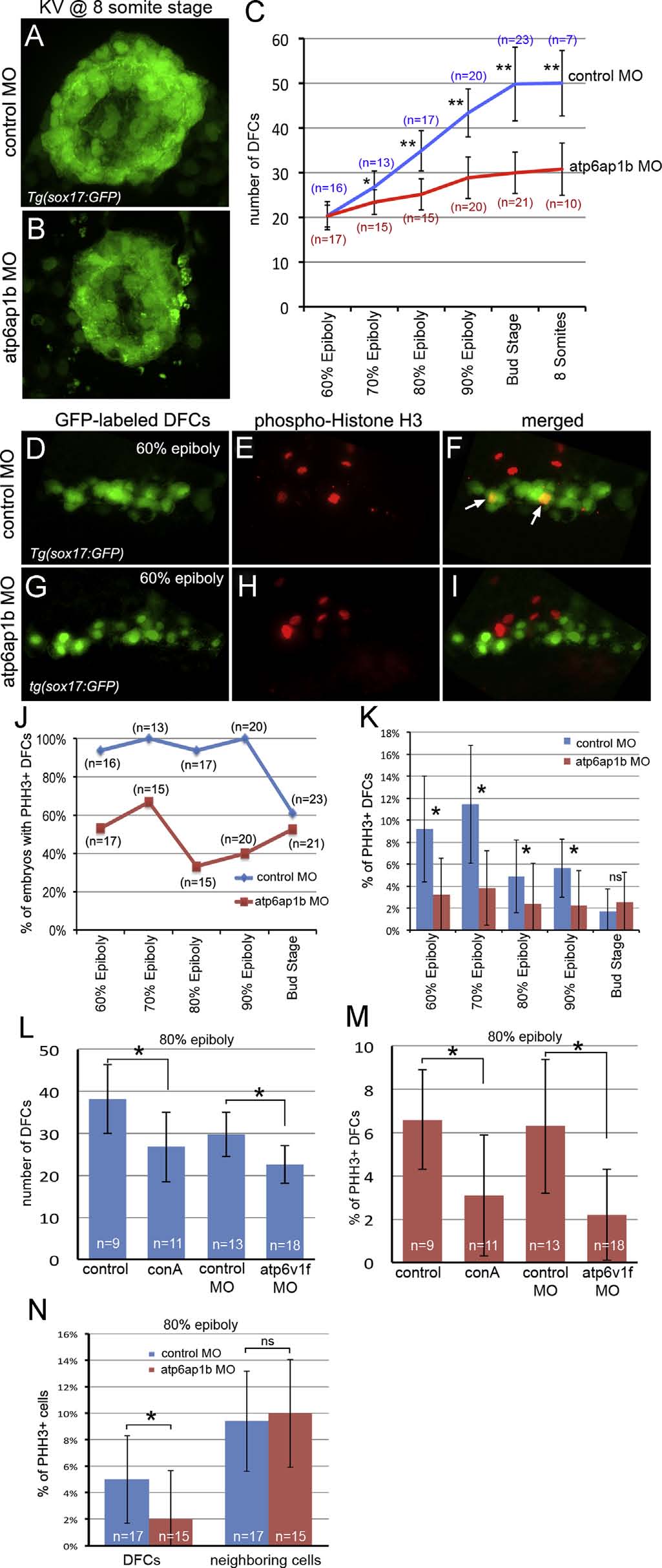
Fig. 8
Atp6ap1b regulates proliferation of DFCs. (A–C) Quantification of DFC/KV cell numbers in Tg(sox17:GFP) embryos between 60% epiboly and 8 somite stages revealed a reduction of cells in atp6ap1b MO injected embryos relative to controls. (D–I) Phosphorylated Histone H3 (pHH3) antibodies labeled proliferating cells in Tg(sox17:GFP) embryos injected with control MO (D–F) or atp6ap1b MO (G–I). Merged images (F,I) of DFCs (green) and pHH3 (red) show examples of proliferating DFCs (arrows) at the 60% epiboly stage. (J) Percentage of embryos with proliferating DFCs at the indicated stages. (K) The percentage of DFCs proliferating in control MO and Atp6ap1 MO injected embryos. (L–M) Embryos injected with atp6v1f MO or treated with concanamycin A between the 50% and 75% epiboly stages were analyzed at the 80% epiboly stage. Treated embryos had a reduced number of DFCs (L) and reduced DFC proliferation (M) as compared to controls. (N) In contrast to DFCs, there was no difference in pHH3 labeling of neighboring dorsal margin cells between atp6ap1b MO and control embryos.
Reprinted from Developmental Biology, 407(1), Gokey, J.J., Dasgupta, A., Amack, J.D., The V-ATPase accessory protein Atp6ap1b mediates dorsal forerunner cell proliferation and left-right asymmetry in zebrafish, 115-30, Copyright (2015) with permission from Elsevier. Full text @ Dev. Biol.