
|
Fig. 6
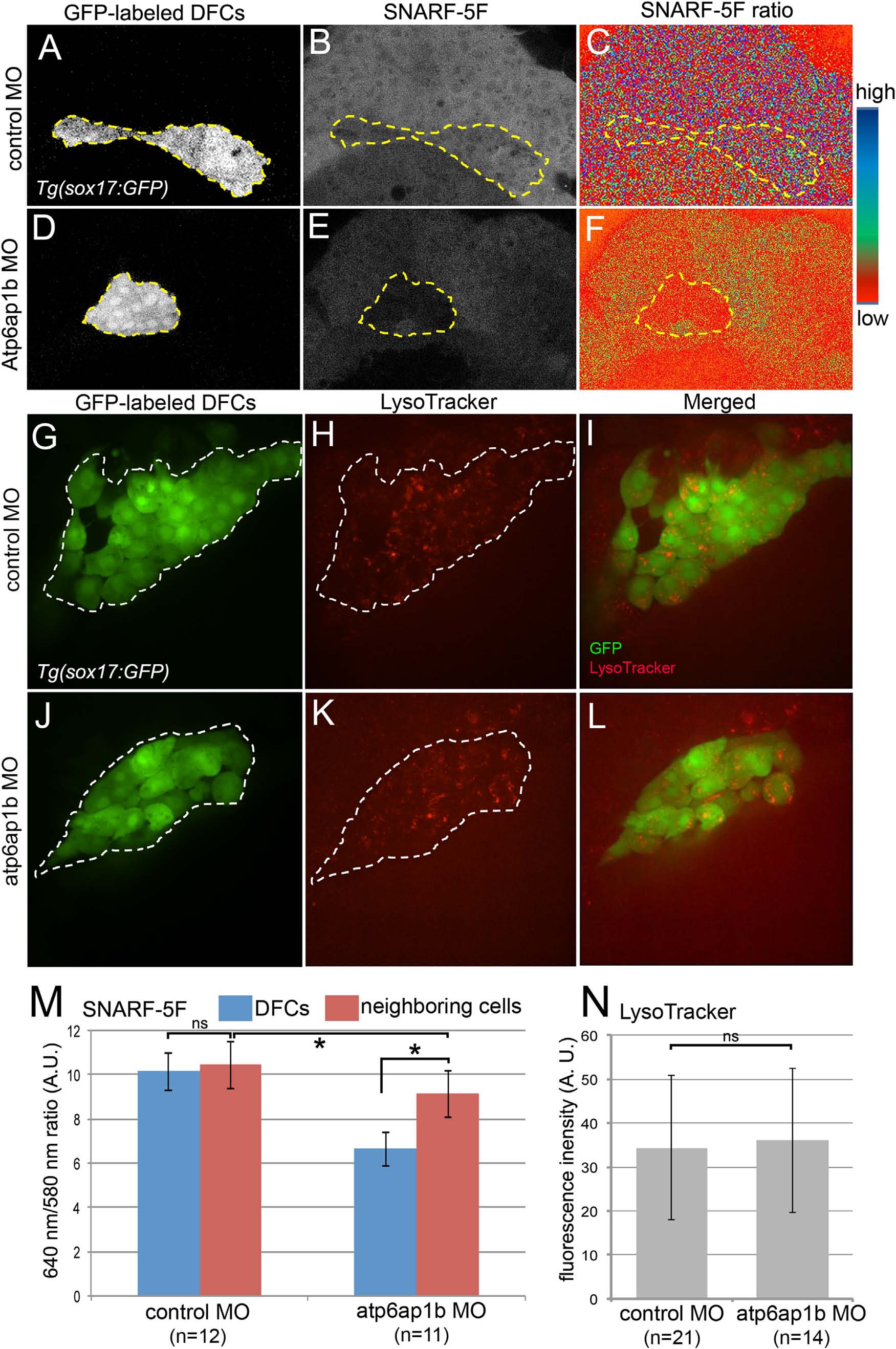
Atp6ap1b depletion reduced cytoplasmic pH of DFCs. (A–F) Analysis of cytoplasmic pH in living embryos during epiboly stages using SNARF-5F fluorescent indicator. DFCs (A,D) are labeled with GFP in Tg(sox17:GFP) embryos and are outlined with a dotted line. (B,E) pH-dependent SNARF-5F fluorescence at 640 nm. (C,F) Pseudocolored ratiometric (640 nm/580 nm) image of SNARF-5F signals. Blue indicates high pH and red indicates low pH. (G–L) LysoTracker vital dye labeling of acidic compartments (e.g. lysosomes) in live embryos. (G,J) GFP+ DFCs are outlined with a dotted line. (H,K) LysoTracker fluorescent staining labels acidic organelles. (I,L) Merged overlay of LysotTracker (red) and DFCs (green). (M) Quantification of SNARF-5F ratio in DFCs and adjacent dorsal margin cells in control MO and atp6ap1 MO injected embryos. (N) Quantification of LysoTracker fluorescence in control and atp6ap1 MO embryos. ns=not significant. AU= arbitrary units.
Reprinted from Developmental Biology, 407(1), Gokey, J.J., Dasgupta, A., Amack, J.D., The V-ATPase accessory protein Atp6ap1b mediates dorsal forerunner cell proliferation and left-right asymmetry in zebrafish, 115-30, Copyright (2015) with permission from Elsevier. Full text @ Dev. Biol.