
Image
|
Figure Caption
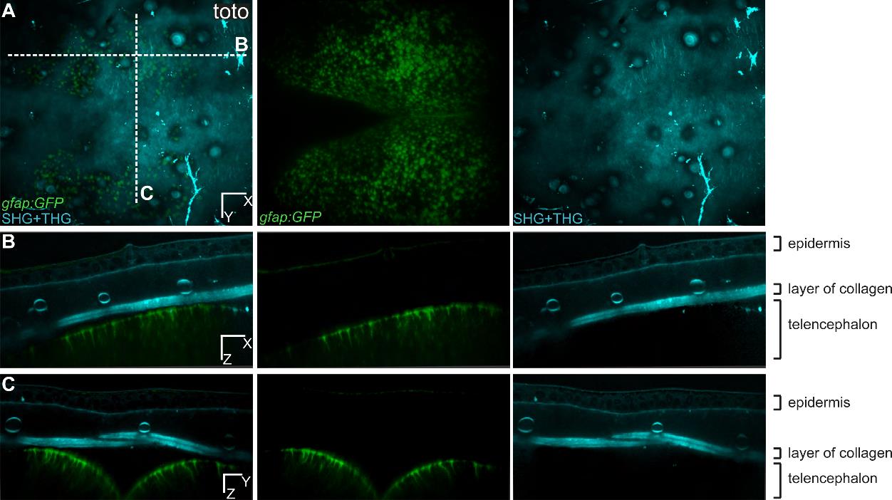
Fig. S4 Multicolor 3D images of RG cells in the pallium and SHG/THG signals of the structures above. Dorsal (A), transversal (B) and cross (C) views for one time point of a transgenic zebrafish gfap:eGFP into a casper background (individual fish named toto). The green channel is showing all the glial cells and the processes are visible (B, C). The cyan channel is showing structures such as collagen, lipids, light-absorbing cells, and the epidermis, where individual cells are visible. The merged panel in (C) is the same as on Fig.1C. Scale bars, 50 µm (A,-C).
Acknowledgments
This image is the copyrighted work of the attributed author or publisher, and
ZFIN has permission only to display this image to its users.
Additional permissions should be obtained from the applicable author or publisher of the image.
Full text @ Development