
|
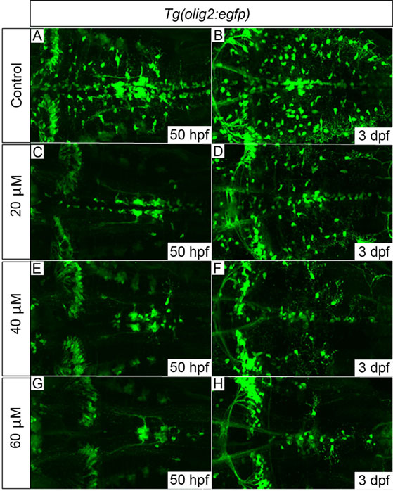
Fig. 7
Tg(olig2:egfp) embryos treated with cyclopamine from 34hpf show dose-dependent OPC proliferation and migration defects. OPCs failed to migrate fully to uniformly populate the hindbrain in cyclopamine-treated embryos (C-H) compared with control embryos (A,B). The development of olig2-positive cerebellar neurons (clearly visible towards the left of panel H) and the abducens motor neurons (axonal projections clearly visible in G) appeared normal, even with the highest dose of 60µM, which suggests that their development is largely Shh-independent. 20 embryos per treatment group; images show dorsal views, anterior to left. Quantification of OPC numbers is shown in supplementary material Fig. S5.