
|
Fig. S4
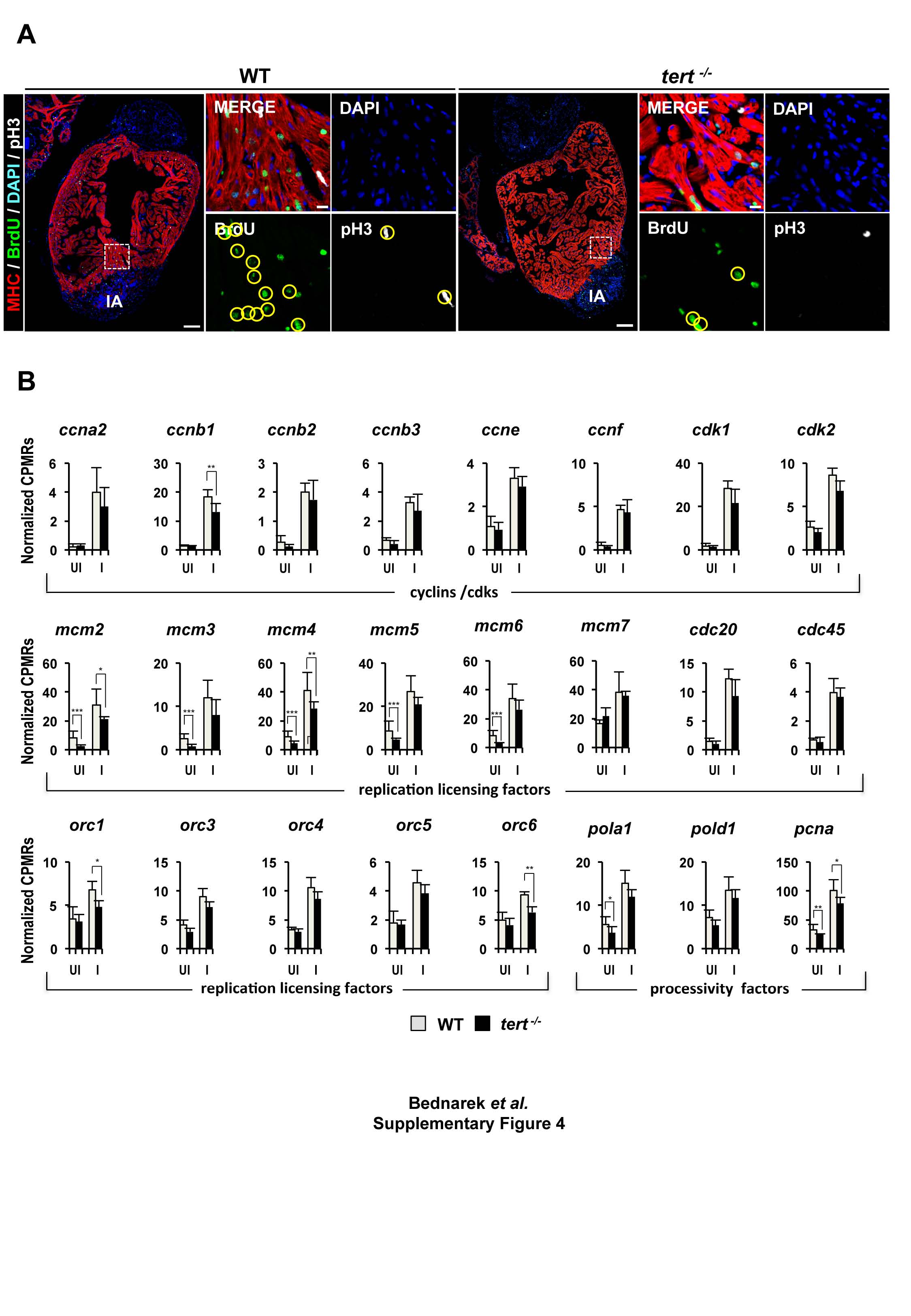
tert loss of function leads to reduced cardiomyoyte proliferation.
(A) Immunofluorescence staining on wildtype (WT) and tert-/- hearts with anti-myosin heavy chain (MHC, red), anti-BrDU (green) and anti-phospho histone 3 (pH3, white). Nuclei are counterstained with DAPI. Boxed areas are shown at higher magnification; yellow circles highlight BrdU/MHC and pH3/MHC double positive cardiomyocytes. Note that fewer pH3 and BrdU-positive cardiomyocytes are found in the tert-/- heart. Scale bars: 100 µm (whole mount views), 10 µm (magnifications).
(B) RNA expression of cyclins, cdks, replication licensing factors and processivity factors implicated in a proliferation response in uninjured and 3 dpi WT and tert-/- hearts. Data are means ± SEM of values obtained from an RNA-seq experiment of 4 biological replicates, each replicate consisting of 3 pooled hearts. CPM, counts per million; dpi, days postinjury. *p<0.05, ** p<0.01, *** p<0.001 (Benjamini-Hochberg adjusted p-values).
Related to Figure 3.