
|
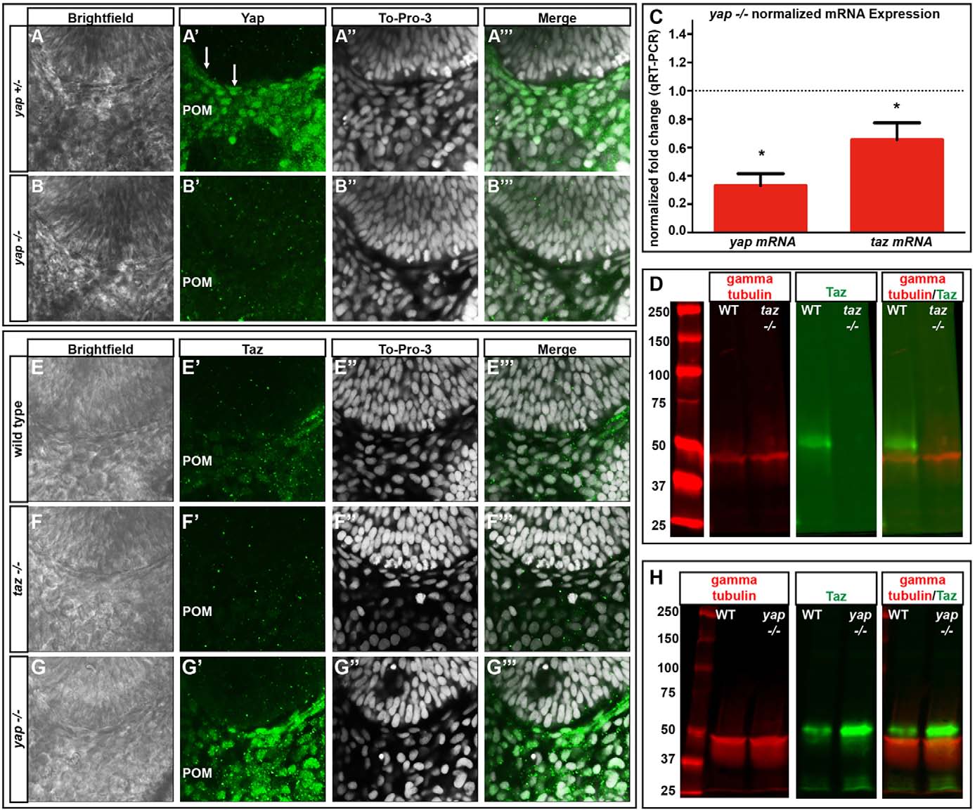
Fig. 3
yap mRNA and Yap protein levels are decreased and Taz protein increased in yap-/- embryos. (A-B′′′) Yap immunoreactivity in wild-type and yap-/- eyes at 28hpf. Yap protein is present in flattened RPE nuclei (arrows) and periocular mesenchyme (POM) in yap+/- embryos, whereas nuclear Yap staining is absent in the yap-/- mutant. (C) qRT-PCR analysis of whole embryos at 32hpf showing a decrease in yap (3-fold, *P=0.0002) and taz (1.5-fold, *P=0.0270) mRNA in yap-/- mutants. Dotted line indicates normalized expression levels of yap and taz in wild-type embryos. An unpaired t-test was performed and statistical significance determined using the Holm-Sidak method. Error bars represent s.e.m. (D) Western blot showing Taz protein (~52 kDa) in wild-type and its absence in taz-/- adult heart tissue. (E-G′′′) Taz immunoreactivity in wild-type, taz-/- and yap-/- embryos at 28hpf. (H) Western blot of Taz protein from 2dpf wild-type (n=20) and yap-/- mutant (n=20) whole embryos.