
Image
|
Figure Caption
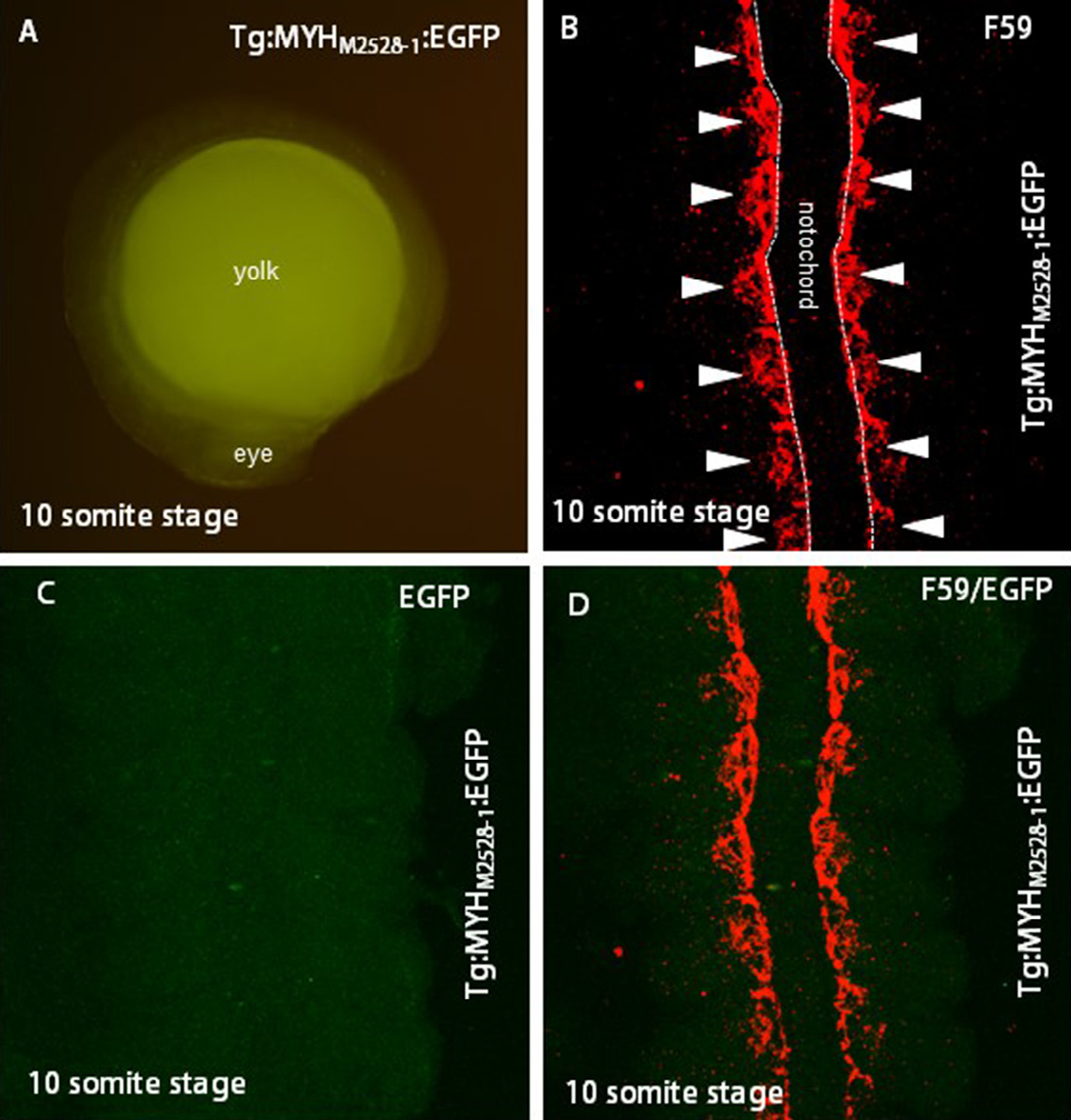
Fig. 7
Adaxial cell-derived slow muscle progenitor in Tg:MYHM2528-1:EGFP transgenic embryo. (A) Ten somite stages of Tg:MYHM2528-1:EGFP transgenic embryo by fluorescent microscope observation. Lateral view. No EGFP expression was observed. (B–D) Immunohistochemistry of Tg:MYHM2528-1:EGFP transgenic embryo at 10 somite stage. Dorsal view. (B) F59 antibody; (C) EGFP; and (D) merged view. Scale bars: 50 µm. In panel B, adaxial cell-derived slow muscle progenitors are indicated by arrowheads.
Acknowledgments
This image is the copyrighted work of the attributed author or publisher, and
ZFIN has permission only to display this image to its users.
Additional permissions should be obtained from the applicable author or publisher of the image.
Reprinted from Mechanisms of Development, 137, Ahammad, A.K., Asaduzzaman, M., Asakawa, S., Watabe, S., Kinoshita, S., Regulation of gene expression mediating indeterminate muscle growth in teleosts, 53-65, Copyright (2015) with permission from Elsevier. Full text @ Mech. Dev.