
|
Fig. 4
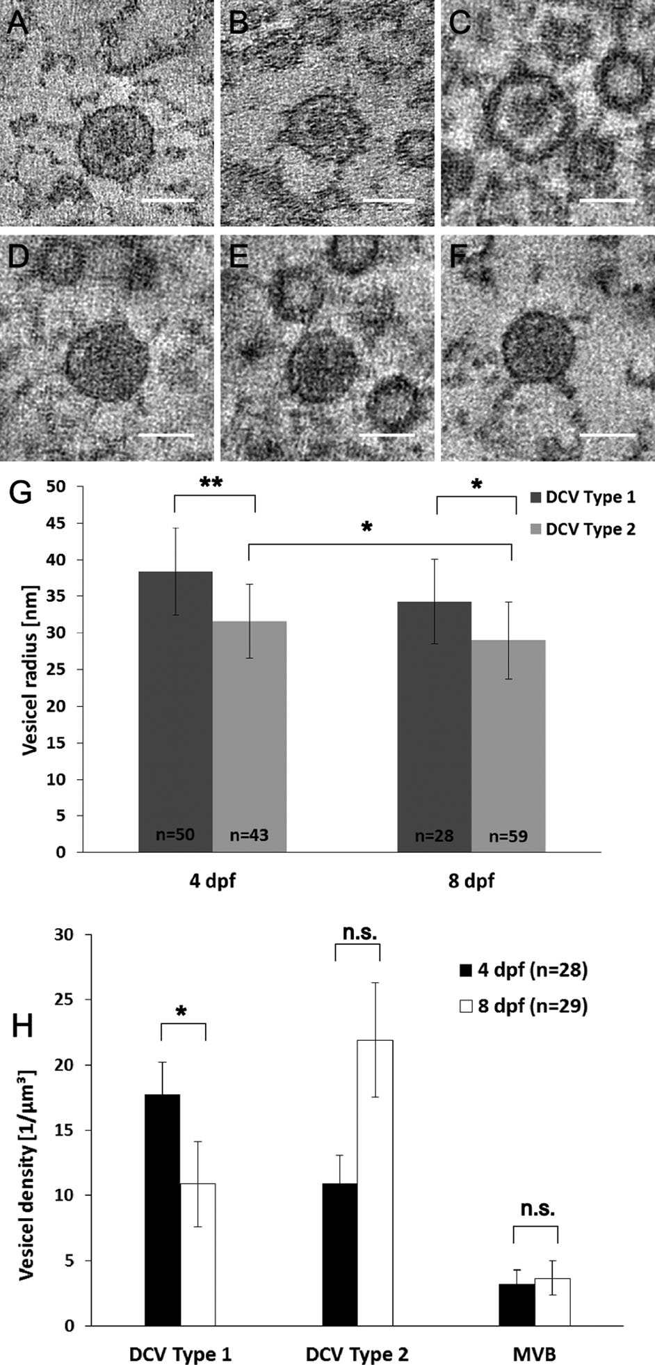
Two types of dense-core vesicles differ in size and appearance. Three different tomogram slices through the middle of type 1 dense-core vesicles (DCV) are shown (A-C). Type 1 DCVs have an electron-dense core surrounded by an electron-clear structure between the membrane and the core. In D-F three different tomogram slices through the middle of type 2 DCVs with a homogeneous dense structure are shown. Type 1 DCV have a radius (G) of 38.4 ± 6.0 nm (n = 50 vesicles from 26 single and double tilt tomograms, mean ± SD) in 4-dpf zebrafish larvae and a mean radius of 34.3 ± 5.8 nm (n = 28 vesicles from 16 single and double tilt tomograms, mean ± SD) in 8-dpf zebrafish larvae, differing significantly [**P < 0.001 (4 dpf) and *P < 0.05 (8 dpf), KS test] from the radius of type 2 DCV of 31.6 ± 5.1 nm (n = 43 vesicles from 18 single and double tilt tomograms, mean ± SD) in 4-dpf zebrafish larvae and 29.0 ± 5.3 nm (n = 59 vesicles from 24 single and double tilt tomograms, mean ± SD) in 8-dpf samples. The density (H) of type 1 dense-core vesicles is significantly higher in NMJs of 4-dpf zebrafish, 17.7 ± 2.5 vesicles/µm3 (n = 28 single and double tilt tomograms, mean ± SEM), than in NMJs of 8-dpf zebrafish, 10.9 ± 3.3 vesicles/µm3 (n = 29 single and double tilt tomograms, mean ± SEM). The density (H) of type 2 DCVs is 11.0 ± 2.1 vesicles/µm3 (n = 28 single and double tilt tomograms, mean ± SEM) in NMJs of 4-dpf zebrafish larvae and 21.9 ± 4.4 vesicles/µm3 (n = 29 single and double tilt tomograms, mean ± SEM) in NMJs of 8-dpf zebrafish larvae. The density of multivesicular bodies does not differ significantly in 4-dpf (3.2 ± 1.1 vesicles/µm3, n = 28 single and double tilt tomograms, mean ± SEM) and 8-dpf (3.7 ± 1.3 vesicles/µm3, n = 29 single and double tilt tomograms, mean ± SEM) larvae. Scale bars = 50 nm.