
|
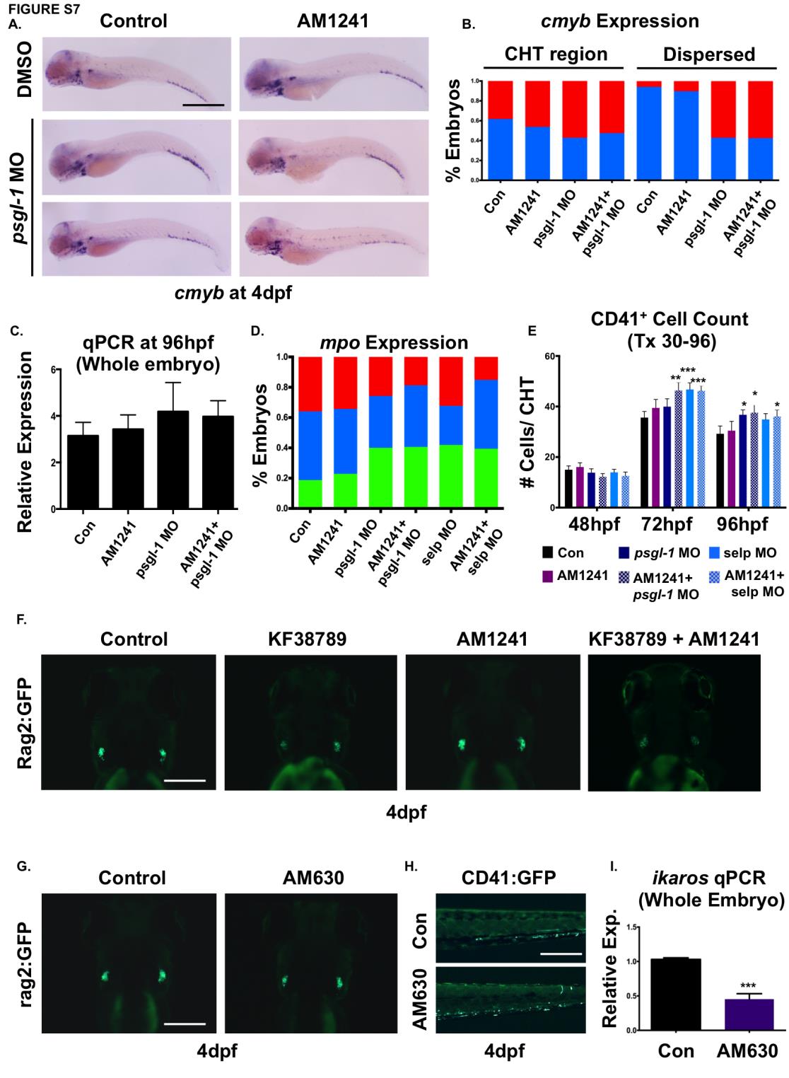
Fig. S7 CNR2-signaling promotes HSPC migration to secondary niches via P-selectin.
(A) In psgl-1 morphant embryos, while thymic colonization is reduced, cmyb expression is observed to accumulate in the CHT (middle panel) and/or appear in isolated cells scatter throughout the embryo (lower panel) in the presence and absence of AM1241-treatment (30-96hpf).
(B) Qualitative phenotypic distribution of cmyb expression within the CHT (left side) or dispersed throughout the embryo (right side) in psgl-1 morphants with and without AM1241-treatment compared to sibling controls (n≥35/condition).
(C) qPCR analysis of psgl-1 morphant embryos showed no significant alterations in cmyb expression in the presence or absence of AM1241- : 1.09-fold, psgl-1 MO/DMSO : 1.33-fold, psgl-1 MO/AM1241 : 1.26-fold; NS, 2-tailed t-test, n=25 pooled embryos/condition x 4 replicates).
(D) Qualitative phenotypic distribution of embryos injected with psgl-1 and selp MOs and subsequently exposed to AM1241 revealed mpo expression, indicative of myeloid commitment, was not increased following P-selectin knockdown (n≥30/condition).
(E) Absolute counts of CD41:GFP+ cells from embryos injected with psgl-1 and selp MOs and exposed to AM1241 (30-96hpf, 5µM) revealed inhibition of P-selectin activity caused prolonged retention of HSCs in the CHT compared to controls (48hpf: Uninjected/DMSO: 15±1.5, Uninjected/AM1241:16.1±1.7, psgl-1 MO/DMSO: 13.8±1.5, psgl-1 MO/AM1241:12.2±1.3, selp MO/DMSO: 14.0±1.2, selp MO/AM1241: 12.6±1.5; 72hpf: Uninjected/DMSO: 35.6±2.4, Uninjected/AM1241:39.4±3.4, psgl-1 MO/DMSO: 39.9±3.1, psgl-1 MO/AM1241:46.4±3.2, selp MO/DMSO: 46.7±2.7, selp MO/AM1241: 46.2±2.0; 96hpf: Uninjected/DMSO: 29.2±3.1, Uninjected/AM1241:30.5±3.7, psgl-1 MO/DMSO: 36.7±1.9, psgl-1 MO/AM1241:37.6±3.3, selp MO/DMSO: 34.9±2.3, selp MO/AM1241: 36.1±2.6; *p≤0.05, **p≤0.01, ***p≤0.001, 2-tailed t-test, n≥10/condition).
(F) Embryos exposed to KF38789 (30-96hpf) confirmed the effects of P-selectin on normal and AM1241- enhanced colonization of Rag2:GFP+ lymphoid progenitors in the thymus at 4dpf (n≥10/condition).
(G) Embryos exposed to AM630 during HSC expansion and thymic colonization (48-96hpf) showed decreased Rag2:GFP intensity in the thymus (n≥10/condition).
(H) In vivo imaging of cd41:gfp embryos (48-96hpf) showed exposure to AM630 cause HSC numbers to remain elevated in the CHT at 96hpf (n≥15/condition).
(I) qPCR analysis confirmed decreased ikaros expression at 96hpf in embryos exposed to AM630 (30- 96hpf) (0.65-fold; ***p≤0.001, 2-tailed t-test, n=25 pooled embryos/condition x 4/replicates).
Scale bars: A =800µm, F,G=175µm, H=200µm.