
|
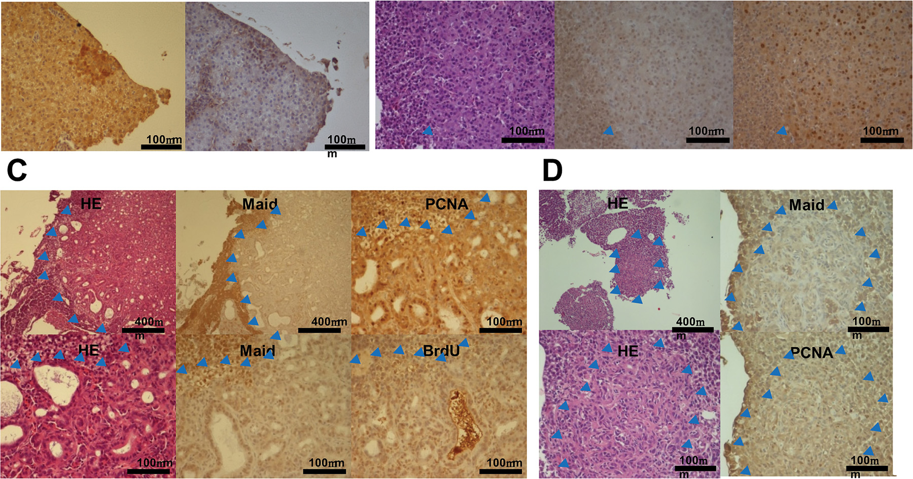
Fig. 5 Immunohistochemical analysis of Maid expression during chemical carcinogenesis in zebrafish.
(A) Liver samples from zebrafish that had been treated for 8 weeks with DEN and subsequently developed preneoplastic foci were subjected to immunohistochemical staining. Results are representative of 11 preneoplastic foci. Red arrow heads indicates Maid overexpressed lesion. Blue arrow heads indicate boundaries of tumor lesions. (B) Liver samples from zebrafish exhibiting HCC after 8 weeks of DEN treatment were subjected toimmunohistochemical staining. Results are representative of 15 HCCs. Blue arrow heads indicate boundaries of tumor lesions. (C) Liver samples from zebrafish exhibiting cholangiocarcinoma after 8 weeks of DEN treatment were subjected to immunohistochemical staining. Results are representative of 4 cholangiocarcinomas. Blue arrow heads indicate boundaries of tumor lesions. (D) Liver samples from zebrafish exhibiting mixed tumors after 8 weeks of DEN treatment were subjected to immunohistochemical staining. Results are representative of 21 mixed tumors. Blue arrow heads indicate boundaries of tumor lesions.