
Image
|
Figure Caption
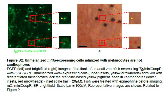
Fig. S2 Unmelanized mitfa-expressing cells admixed with melanocytes are not xanthophores
EGFP (left) and brightfield (right) images of the flank of an adult zebrafish expressing Tg(miniCoopRmitfa: nlsEGFP). Unmelanized mitfa-expressing cells (upper insets, yellow arrowheads) admixed with differentiated melanocytes lack the pteridine-based yellow pigment seen in xanthophores (lower insets, red arrowheads) (inset scale bar = 20µM). Fish were treated with epinephrine before imaging. mC, miniCoopR; BF, brightfield. Scale bar = 100µM. Representative images are shown. Related to Figure 2.
Acknowledgments
This image is the copyrighted work of the attributed author or publisher, and
ZFIN has permission only to display this image to its users.
Additional permissions should be obtained from the applicable author or publisher of the image.
Reprinted from Developmental Cell, 33(6), Iyengar, S., Kasheta, M., Ceol, C.J., Poised Regeneration of Zebrafish Melanocytes Involves Direct Differentiation and Concurrent Replenishment of Tissue-Resident Progenitor Cells, 631-43, Copyright (2015) with permission from Elsevier. Full text @ Dev. Cell