
|
Fig. 2
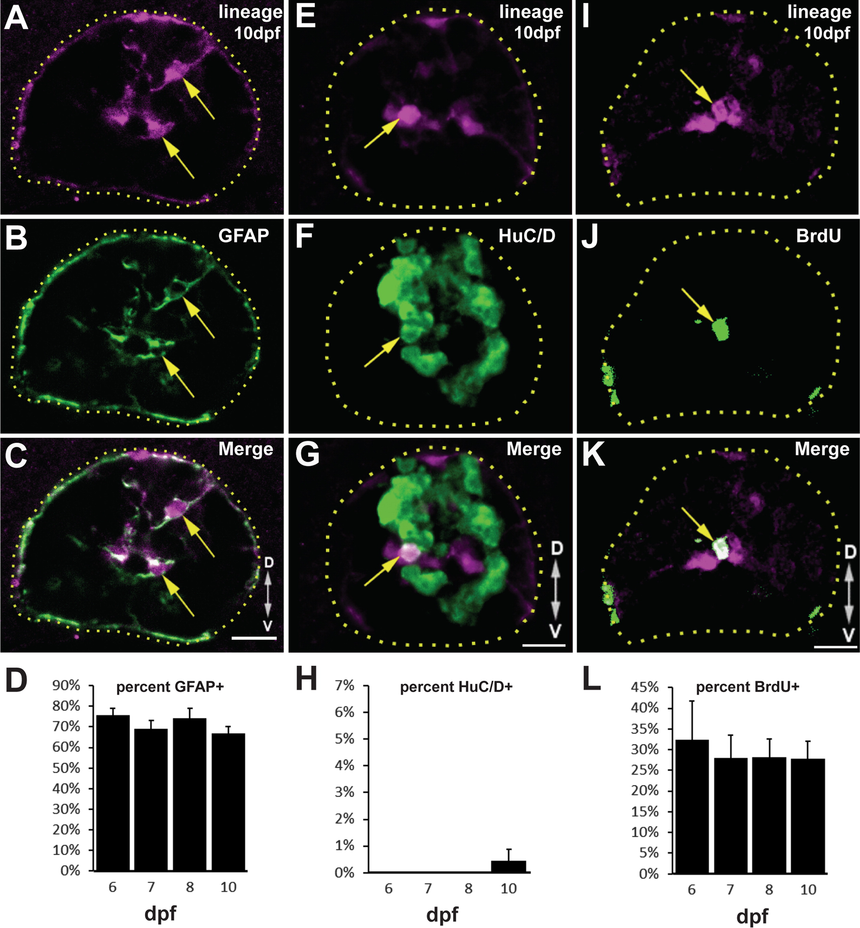
Tg(gfap:CreERT2,CY)zd16 labels a quiescent population of radial glia. (A–D) Following 4-OHT treatment, most mCherry+ cells remain GFAP+ (arrows) through 10 dpf. (E–H) In contrast, very few mCherry+ cells express HuC/D (arrow) beginning at 10 dpf. (I–L) After incubation in BrdU from 4 to 5 dpf, there is no significant expansion of the BrdU-labeled subset of mCherry+ cells (arrow). All images are single confocal slices of 12 µM transverse cryosections. n=25 sections from 5 animals at each timepoint; error bars=SEM; scalebar=10 µM, dotted lines outline the spinal cord, and D/V arrows indicate dorsal and ventral, respectively.
Reprinted from Developmental Biology, 403(1), Briona, L.K., Poulain, F.E., Mosimann, C., Dorsky, R.I., Wnt/ß-catenin signaling Is required for radial glial neurogenesis following spinal cord injury, 15-21, Copyright (2015) with permission from Elsevier. Full text @ Dev. Biol.