
Image
|
Figure Caption
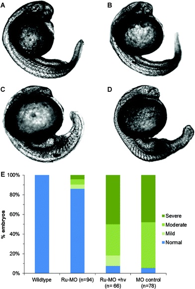
Fig. 5 Representative confocal micrographs of 24–28 hpf zebrafish embryos, showing chd knockdown phenotypes depending on experimental protocol. (A) Wildtype embryo, uninjected. (B) Ru-MO-chd, incubated in the dark, showing normal development. (C) Ru-MO-chd, irradiated for 5 min at 1 hpf with 450 nm light, showing chd knockdown phenotype. (D) Bis-azido MO-chd showing chd knockdown phenotype. All embryos were injected at 1-cell stage and imaged with a 10× air objective (Olympus UPlanSApo, NA = 0.40). (E) Percentage of embryos showing wildtype or degree of phenotypic response.
Figure Data
Acknowledgments
This image is the copyrighted work of the attributed author or publisher, and
ZFIN has permission only to display this image to its users.
Additional permissions should be obtained from the applicable author or publisher of the image.
Full text @ Chem Sci