
|
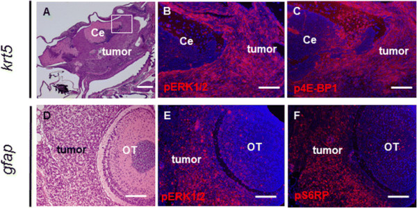
Fig. 4
Activation of the Ras and mTOR pathways in brain tumors. (A) A 6-month-old krt5-derived brain tumor infiltrating both the ventral brain and the VZ. Tumor cells exhibited prominent expression of phospho-ERK1/2 (B) and phospho-4E-BP1 (C). (B) and (C) show immunofluorescence staining of the white framed region in (A). Note the relatively normal cerebellum exhibited much less staining for the two antibodies. (D) A 12-month-old gfap-derived brain tumor showed tumor cell infiltration of the VZ surrounding the OT. Tumor cells exhibited prominent expression of pERK1/2 (E) and pS6RP (F). Note that paraffin sections were used for immunofluorescence. Ce, cerebellum; OT, Optic tectum. Scale bars, 200 µm for A; 40 µm for B, C, D, and E.