
|
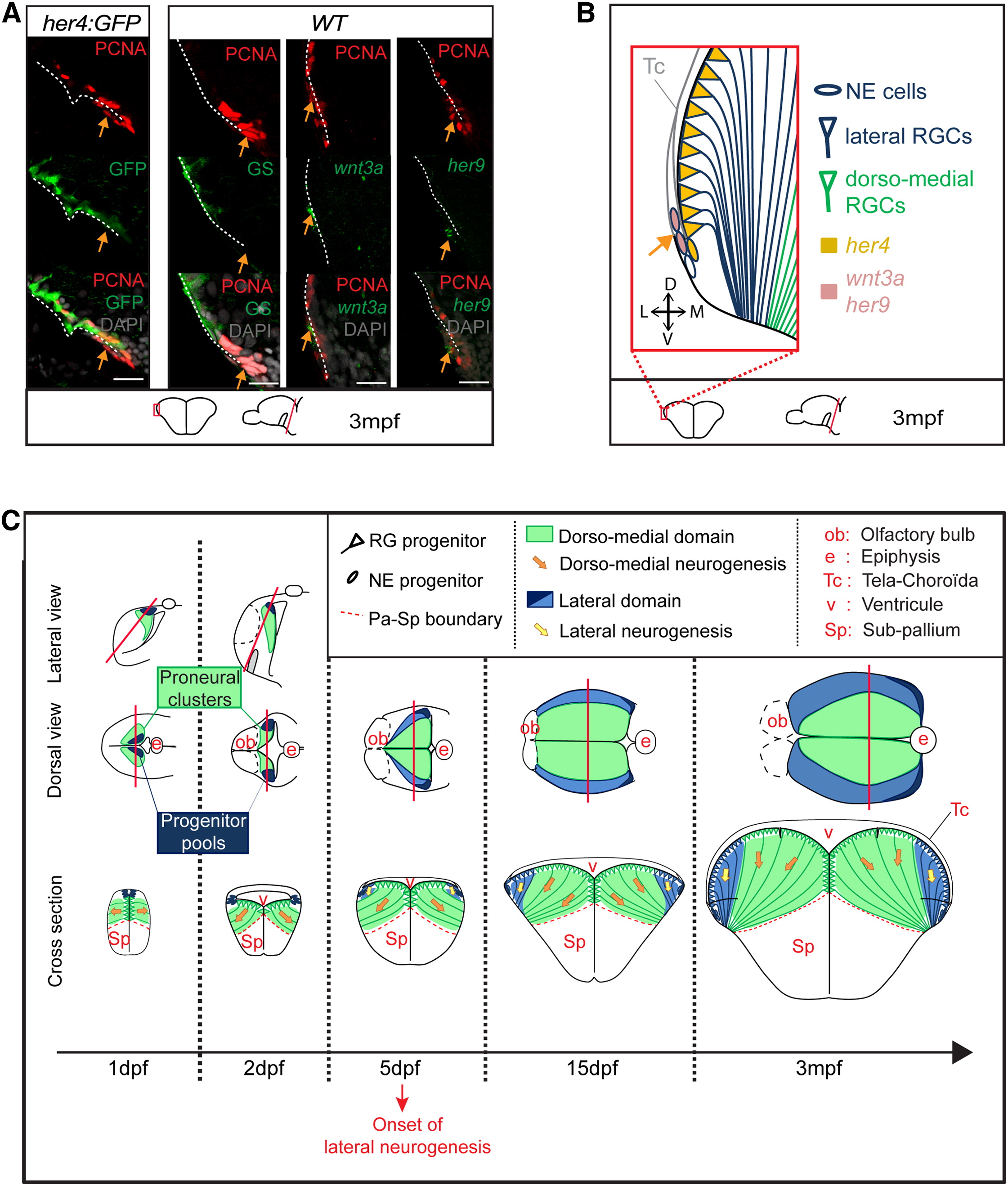
Fig. 7
Dual Embryonic Origin of Pallial aNSCs and Persistence of Adult NE Cells
(A) Posterior cross-sections showing the lateral edge of the adult VZ in her4:GFP fish (left) or WT fish (right) stained as indicated. Arrows point to the her4/PCNA+ NE pool of progenitors.
(B) Scheme of the lateral edge of the adult posterior pallial VZ (as shown in A) depicting RGCs (blue triangles), NE cells (blue), her4 expression (yellow), and wnt3a/her9 expression (pink). The her4 NE pool contains wnt3a+ and/or her9+ proliferating progenitors at the junction between the tela-choroïdea and the posterior pallial VZ edge.
(C) Summary of pallium formation: the dorsomedial pallial NSCs and neurons (green) derive from embryonic neurogenic progenitors. The lateral pallial NSCs and neurons (blue) derive from few NE “progenitor pool” (dark blue) that are first amplified and become neurogenic only from 5 dpf onward and persist lifelong. Both aNSC populations remain segregated in space.
See also Figure S7.
Reprinted from Developmental Cell, 30(2), Dirian, L., Galant, S., Coolen, M., Chen, W., Bedu, S., Houart, C., Bally-Cuif, L., Foucher, I., Spatial Regionalization and Heterochrony in the Formation of Adult Pallial Neural Stem Cells, 123-36, Copyright (2014) with permission from Elsevier. Full text @ Dev. Cell