
|
Fig. 6
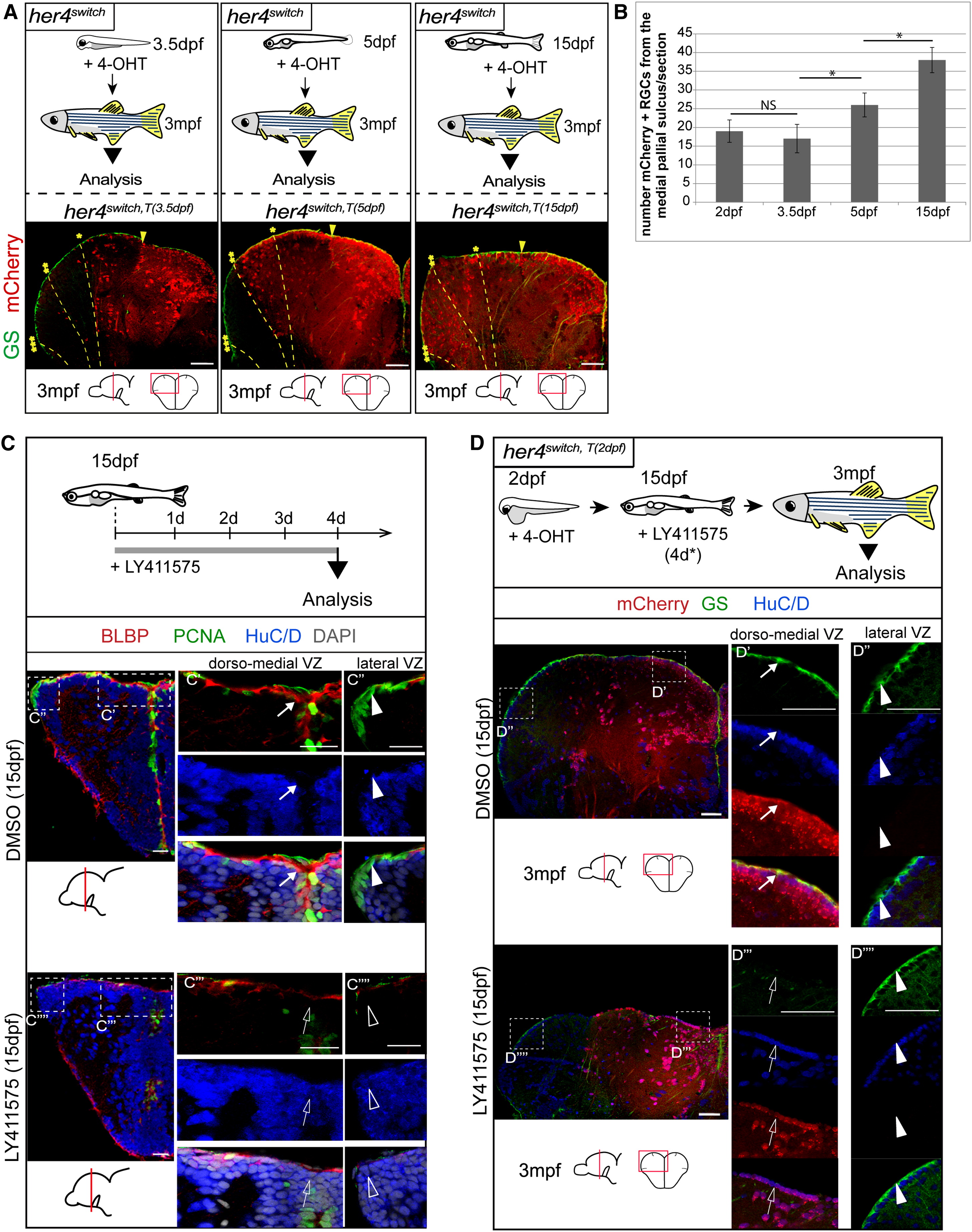
Lateral Progenitors Progressively Express her4 and Become Notch-Sensitive at Juvenile Stages but Maintain a Cryptic Boundary with the Dorsomedial VZ
(A) Adult fate of progenitors expressing her4 at 3.5 dpf (her4switchT(3.5 dpf)), 5 dpf (her4switchT(5 dpf)), or 15 dpf (her4switch,T(15 dpf)): experimental design and respective cross-sections of adult telencephali immunostained as indicated. Yellow stars and dotted lines indicate the mCherry+ boundary observed after recombination at different stages (single star: recombination at 2 dpf; double stars: recombination at 5 dpf; triple stars: recombination at 15 dpf). Yellow arrowhead points to the sulcus ypsiloniformis.
(B) Numbers of mCherry+ RGCs after recombination at 2 dpf, 3.5 dpf, 5 dpf, and 15 dpf in her4switch fish counted from the sulcus ypsiloniformis up to the lateral edge of the VZ. mCherry+/GS+ RGCs comprise 33% of the VZ at 3.5 dpf, and 85% after a recombination at 15 dpf. Values are presented as mean ± 95%CI (ANOVA, p < 0.05).
(C) Notch sensitivity of pallial progenitors at juvenile stages. Experimental design and cross-sections of the telencephalon in control (top) or LY411575-treated fish (bottom) immunostained as indicated. Magnification of the dorsomedial VZ (C′ and C′′′) and lateral VZ (C′′ and C′′′′), immunostained as indicated. Arrows point to medial progenitors, arrowheads to lateral progenitors, empty arrows/arrowheads indicate when these progenitors are depleted.
(D) Adult analysis of the telencephalon of fish treated with a Notch inhibitor at juvenile stage. Experimental design and cross-sections of the telencephalon in control (top) or LY411575-treated fish (bottom) immunostained as indicated. Arrows point to medial (mCherry+) progenitors, arrowheads to lateral (mCherry) progenitors, empty arrows/arrowheads indicate when these progenitors are depleted.
See also Figure S6.
Reprinted from Developmental Cell, 30(2), Dirian, L., Galant, S., Coolen, M., Chen, W., Bedu, S., Houart, C., Bally-Cuif, L., Foucher, I., Spatial Regionalization and Heterochrony in the Formation of Adult Pallial Neural Stem Cells, 123-36, Copyright (2014) with permission from Elsevier. Full text @ Dev. Cell