
|
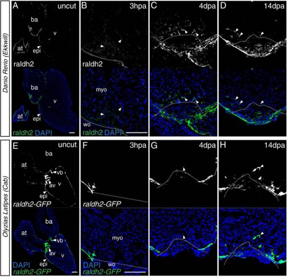
Fig. 4
Raldh2 analysis after ventricular resection. A–D: Fluorescent images of immunostaining for raldh2 in the zebrafish heart (black-white images in the upper row). Raldh2 localization was observed in the epicardium of the uncut heart (A). At 3 hpa (hours post-amputation), endocardial localization of raldh2 was observed (B). Endocardial localization was strengthened and retained at 4 dpa (C) and 14 dpa (D). Raldh2 localization was also observed at the wound site at 4 dpa (C) and 14 dpa (D). E–H: Fluorescent images of heart of the medaka raldh2-GFP transgenic line (black-white images in the upper row). Raldh2 expression was observed in the epicardium of the uncut heart (E). At 3 hpa, only epicardial expression was observed (F). Raldh2-GFP positive fibroblast-like cells were observed in the wound and myocardial area at 4 dpa (G) and 14 dpa (H), whereas no endocardial raldh2 expression was observed. Arrowheads indicate the raldh2 expression or localization. The dashed line indicates the approximate injury border. at, atrium; av, atrio-ventricular valves; ba, bulbus arteriosus; epi, epicardium; myo, myocardium; v, ventricle; vb, ventriculo-bulbal valves; wo, wound area. Shown are representative images from experiments repeated > 3 times. Scale bars = 100 µm.