
|
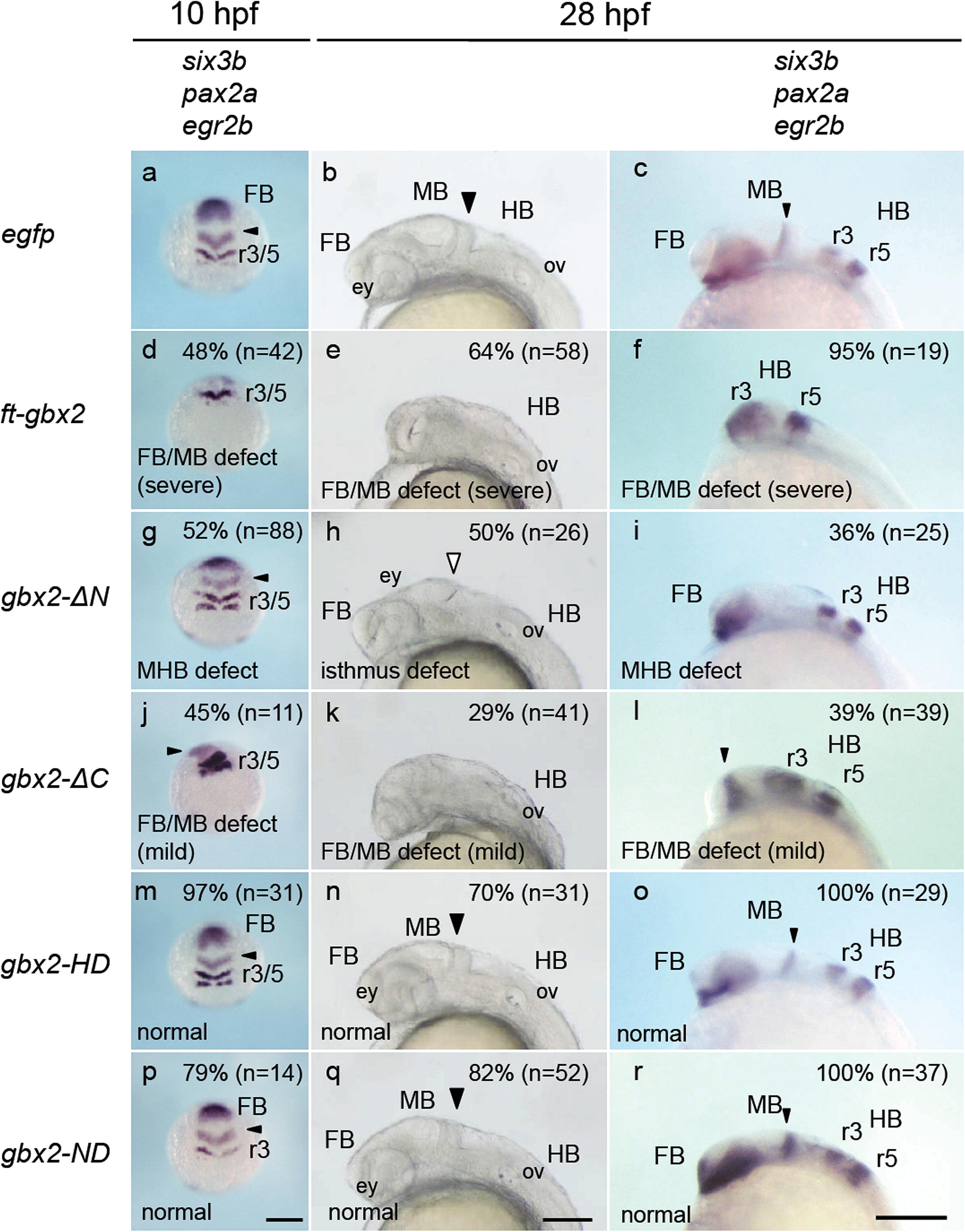
Fig. 9
Effects of gbx2 deletion mutants on brain formation. Embryos were injected with mRNA for egfp, ft-gbx2, gbx2-ΔN, gbx2-ΔC, gbx2-HD, or gbx2-ND and examined at the bud stage (10 hpf, left column) and 28 hpf (right column) for the expression of six3b in the forebrain, pax2a at the MHB, and egr2b in rhombomeres 3 and 5 (r3 and r5, respectively) of the hindbrain or for the morphology of live embryos at 28 hpf (middle column). The most striking phenotypes and their proportions are shown in the respective panels. The positions of the MHB are indicated by small triangles. Normal and disrupted isthmuses are shown with solid and open large triangles, respectively. ey, eye; FB, forebrain; HB, hindbrain; MB, midbrain; ov, otic vesicle. Scale bars, 200 µm.
Reprinted from Mechanisms of Development, 130(11-12), Nakayama, Y., Kikuta, H., Kanai, M., Yoshikawa, K., Kawamura, A., Kobayashi, K., Wang, Z., Khan, A., Kawakami, K., and Yamasu, K., Gbx2 functions as a transcriptional repressor to regulate the specification and morphogenesis of the mid-hindbrain junction in a dosage- and stage-dependent manner, 532-52, Copyright (2013) with permission from Elsevier. Full text @ Mech. Dev.