Fig. 3
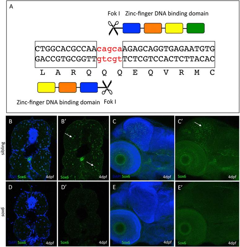
Targeted mutagenesis of the zebrafishsox6locus. (A) Schematic representation of the zinc-finger nuclease designed to target genomic sequences in exon 8, upstream of the HMG box, of the zebrafish sox6 gene. (B-E) Immunohistochemical detection of Sox6 protein using a Sox6-specific antibody; Sox6 protein is detectable in the nuclei of fast muscle fibers (arrows) (B and B′) and the optic tectum (arrow) (C and C′) of wild-type embryos. In sox6 homozygous mutants, by contrast, no signal is detected in either the muscle (D and D′) or the optic tectum (E and E′), indicating the successful generation of a null mutant. (Blue signal = DAPI).

