
|
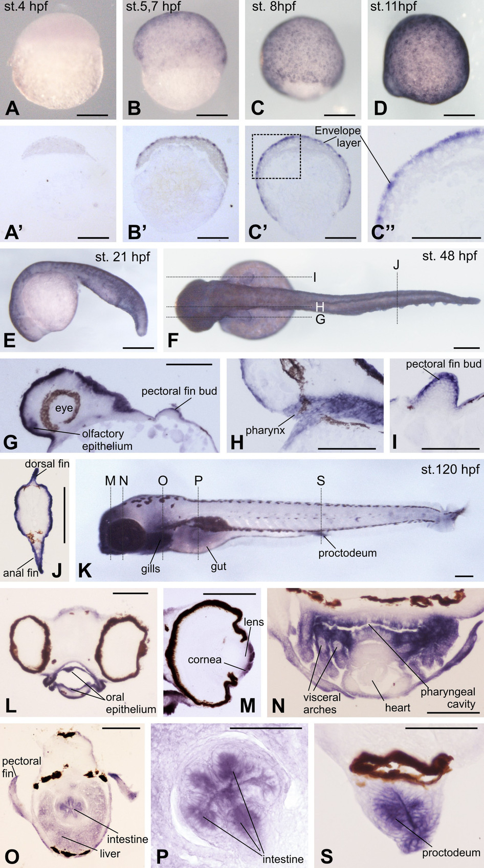
Fig. 2
Spatial expression pattern of DAg1 in Danio rerio embryos as revealed by in situ hybridization.
(A–D and E). Up to 21hpf, DAg1 is expressed only in the superficial enveloping layer. (A′–C′). Histological sections of embryos shown on A–C. (C′′). Enlarged fragment framed on C′ by dotted line. (F–J). Dorsal view of the embryo at hatching stage (F) and histological sections (G–J) of sibling embryos corresponding to the planes indicated by dotted lines on F. (K–S). Left side view of embryo at 5 days stage (K) and histological sections (L–S)of sibling embryos corresponding to the planes indicated by dotted lines on K. Scale bar everywhere is 200µm.