
|
Fig. 6
Actin Polymerization Activity of Fmnl3 Is Autoinhibited by Intramolecular Interaction between Its N-Terminal and C-Terminal Regions
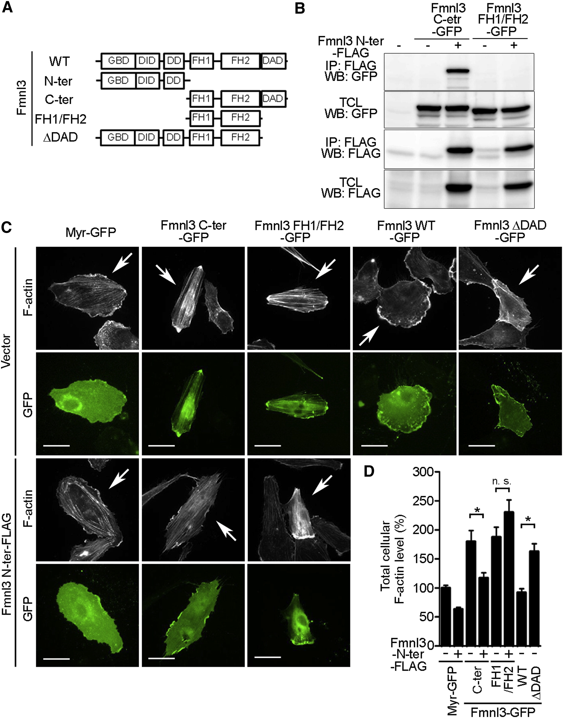
(A) Schematic illustrations of Fmnl3 and its mutants. DID, diaphanous inhibitory domain; DD, dimerization domain; ter, terminus.
(B) 293T cells were transfected without () or with (+) the plasmid encoding Fmnl3 N-ter-FLAG together with either the plasmid expressing Fmnl3 C-ter-GFP or that encoding Fmnl3 FH1/FH2-GFP as indicated at the top. Immunoprecipitates (IP: FLAG) of cell lysates and total cell lysate (TCL) aliquots were subjected to western blot (WB) analyses with anti-GFP and anti-FLAG antibodies as indicated on the left.
(C) HUVECs transfected with the plasmid encoding Myr-GFP (0.5 µg) or that encoding C-terminally GFP-tagged Fmnl3 (0.5 µg) or its mutants (0.5 µg, Fmnl3 C-ter-GFP, Fmnl3 FH1/FH2-GFP, and Fmnl3 ΔDAD-GFP), as indicated at the top, together with the empty vector (1 µg, Vector) or the plasmid expressing C-terminally FLAG-tagged Fmnl3 N-ter mutant (1 µg, Fmnl3 N-ter-FLAG), as indicated on the left, were stained with rhodamine-phalloidin. The rhodamine (F-actin) and GFP images are shown. Arrows indicate GFP signal-positive cells. Scale bars, 30 µm. Note that expression of either Fmnl3 C-ter-GFP or Fmnl3 FH1/FH2-GFP induced stress fiber formation, while thin actin fibers were formed throughout the cell in response to expression of Fmnl3 ΔDAD-GFP.
(D) Total cellular F-actin levels as observed in (C) were quantified by measuring the rhodamine fluorescence intensity of individual cells and expressed as percentages relative to that observed in Myr-GFP-expressing cells. Data are shown as means ± SEM (n = 17). p < 0.05; n.s., no significance.
See also Figure S6.
Reprinted from Developmental Cell, 32, Wakayama, Y., Fukuhara, S., Ando, K., Matsuda, M., Mochizuki, N., Cdc42 Mediates Bmp-Induced Sprouting Angiogenesis through Fmnl3-Driven Assembly of Endothelial Filopodia in Zebrafish, 109-22, Copyright (2015) with permission from Elsevier. Full text @ Dev. Cell