
|
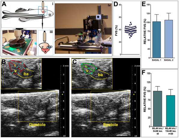
Fig. 1
Echocardiographic image acquisition and basal fractional volume shortening (FVS) quantification.
(A) Schematic representation of animal positioning for image acquisition and picture of the set up. (i) Animals are positioned ventrally and (ii) are immobilized in the same way as for surgical procedures, in a Petri dish, and are covered with fish water containing anaesthetic solution. This positioning allows for a transducer to be placed directly over the body wall at the level of the heart (iii). The transducer is attached to a holder to allow a stable position during acquisition (iv). (B,C) Details from representative 2D echocardiography images from an uninjured zebrafish heart showing maximal ventricular dilatation (B, diastole) and maximal ventricular contraction (C, systole). The diastolic (red) and systolic (green) ventricular areas are outlined and the length of the apical image long axis is also indicated (L). Red and green lines in B and C highlight ventricular border in diastole and systole, respectively. Yellow lines indicate the bulbus arteriosus (BA). (D) FVS obtained in basal conditions (n = 47, mean ± SD = 39 ± 5). (E) Comparison of FVS in basal conditions at two different days, with an interval of 7 days. Shown are means ± SD. The relative FVS (RFVS) of BASAL2 versus BASAL1 within the same animal are statistically comparable (p = 0.1099, Wilcoxon matched-pairs signed rank test). (F) FVS measured in basal conditions with different dosages of anesthesia and throughout time in the same animal. Initial anesthesia conditions are the same as for all acquisitions (60 µM tricaine/3 mM isoflurane). The final acquisition was taken 20 minutes later and the final anesthesia dose was 60 µM tricaine/15 mM isoflurane. Differences in the average FVS are not statistically significant (p = 0.1094, Wilcoxon matched-pairs signed rank test). A, atrium; ba, bulbus arteriosus; FVS, fractional volume shortening; L, length of the apical image long axis; RFVS, relative fractional volume shortening; v, ventricle.