
|
Fig. 9
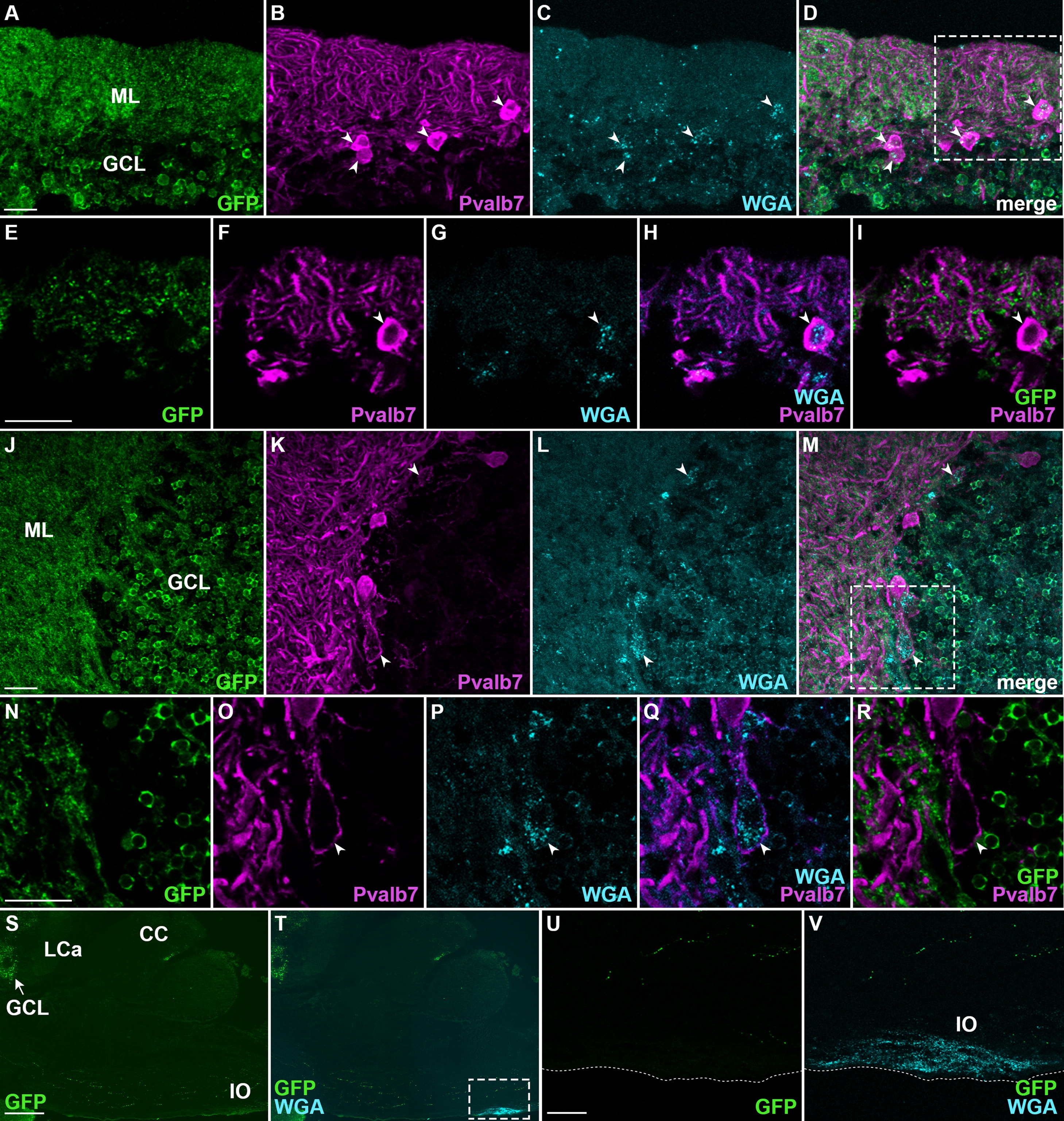
Detection of cerebellar neural circuitry with WGA tracing. The gSA2AzGFF152B; UAS:GFP line was crossed with UAS:AcGFP-P2A-WGA. Sagittal sections of the adult cerebellum were stained with anti-GFP (green), anti-Pvalb7 (magenta), and anti-WGA (cyan) antibodies. (A-D) and (J-M) represent two different areas of the ML/GCL boundary of the cerebellum. Expression of GFP (A, J), Pvalb7 (B, K), and WGA (C, L), and merged images (D, M) are shown. Projection views of histological sections. (E-I) High magnification views of the box in D. (N-R) High magnification views of the box in M. Optical sections (E-I, N-R). Pvalb7+ Purkinje cells that incorporated WGA are indicated by arrowheads (B-D, F-I). Eurydendroid cell with a Pvalb7 soma, that received Pvalb7+ axon(s) and incorporated WGA, is indicated by arrowheads (K-M, O-R). Note both Purkinje and eurydendroid cells are GFP-negative. (S, T) Low magnification views of the hindbrain. Expression of GFP (S-V) and WGA (T, V). (U, V) High magnification views of the box in T (IO region). The ventral bottoms of the hindbrain are indicated by dotted lines (U, V). Note that IO neurons incorporated WGA but GFP-negative. Scale bars: 20 µm in A (applied to A-D); 20 µm in E (applied to E-I); 20 µm in J (applied to J-M); 20 µm in N (applied to N-R); 200 µm in S (applied to S, T); 40 µm in U (applied to U, V).
Reprinted from Developmental Biology, 397(1), Takeuchi, M., Matsuda, K., Yamaguchi, S., Asakawa, K., Miyasaka, N., Lal, P., Yoshihara, Y., Koga, A., Kawakami, K., Shimizu, T., Hibi, M., Establishment of Gal4 transgenic zebrafish lines for analysis of development of cerebellar neural circuitry, 1-17, Copyright (2015) with permission from Elsevier. Full text @ Dev. Biol.