
|
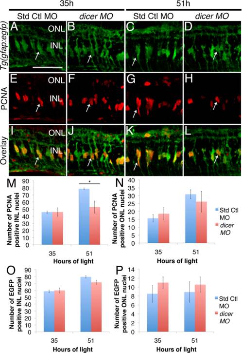
Fig. 3 Dicer knockdown does not affect Müller glia proliferation in light-damaged retinas. Adult albino Tg(gfap:egfp)nt11 zebrafish were intravitreally injected and electroporated with standard control or dicer morpholino prior to the start of light damage. A–L: EGFP-positive Müller glia (arrows) were observed in comparable numbers between dicer and standard control morphants at all time points (A–D, I–L, O, P). E,F: PCNA-positive INL cells were observed as mainly single nuclei in dicer and standard control morphant retinas at 35h of light damage (arrows). IJ: Overlay of these images revealed that many Müller glia co-labeled with PCNA as they re-entered the cell cycle. G: Standard control morphant retinas at 51h of light damage exhibited doublets or early columns of PCNA-positive INL cells. H: Single PCNA-positive INL cells rather than doublets predominated in dicer morphant retinas (arrows). M–N: Differences in proliferation in dicer morphants resulted in significantly fewer PCNA-positive INL cells at 51h of light, but no difference in PCNA-positive ONL cells. Std Ctl MO, Standard Control morphant; dicer MO, dicer morphant; INL, inner nuclear layer; ONL, outer nuclear layer. Scale bar in A = 50 µm and applies to B–L; *P<0.05 using two-way ANOVA with a Tukey′s post-hoc test, n=7.