
|
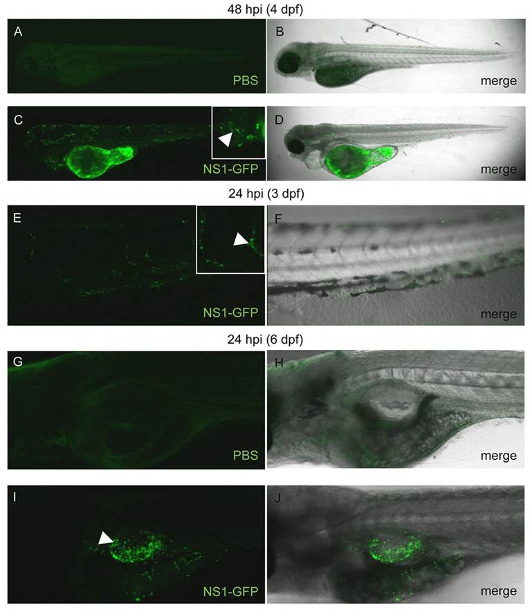
Fig. 5 NS1-GFP shows infection of the zebrafish cardiovascular system and swimbladder. (A–D) Single focal planes of PBS- or NS1-GFP-injected, fixed casper mutant zebrafish at 48 hpi, side mounted, anterior left, dorsal top, ×4 magnification. (A) Fluorescence micrograph of a PBS-injected control showing background autofluorescence. The data presented represent three individual experiments, a mean of n=6 fish per treatment. (B) Panel of PBS control showing merged fluorescence and brightfield micrographs. (C) Fluorescence micrograph of NS1-GFP-infected zebrafish. Punctate fluorescence confirms viral replication. Inset is a computer magnification of the pericardial region. White arrowhead points to fluorescence in the heart. (D) Merge of NS1-GFP-infected embryo fluorescence and brightfield micrographs. (E,F) 3D reconstruction of NS1-GFP-infected, live zebrafish, 24 hpi, side mounted, anterior left, dorsal top, magnification ×10. Punctate fluorescence was observed in major blood vessels. Inset is a computer zoom of two infected blood vessels. White arrowhead denotes fluorescence in an intersomitic blood vessel. (G–J) 3D reconstruction of casper mutant zebrafish after injection of either PBS or NS1-GFP into the swim bladder at 5 dpf, fixed at 24 hpi, side mounted, anterior left, dorsal top. (G) Fluorescence micrograph of zebrafish with PBS injected into the swim bladder, the image shows background autofluorescence. The data represent two individual experiments, a mean of n=5 fish per treatment. (H) Merged panel of PBS-injected control fluorescence and brightfield micrographs. (I) Fluorescence micrograph of NS1-GFP swim bladder infection. Green punctate fluorescence is likely to show infection of epithelial cells around the swim bladder. (J) Fluorescence and brightfield micrograph merge for NS1-GFP swim bladder infection. Infection with the NS1-GFP reporter virus in transparent zebrafish larva allows for visualization of viral replication, spread, resolution of infection and cell tropism in vivo.