
|
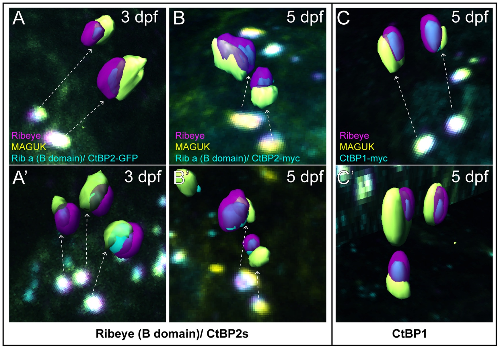
Fig. 6 Ribeye B-domain/CtBP2s and CtBP1 localizes to the basal end of synaptic ribbons facing the postsynaptic density.
Isosurface renderings of ribbon synapses extrapolated from z-stack confocal images of Ribeye b (magenta), GFP or myc (cyan), and MAGUK (yellow). Dashed arrows indicate the ribbon synapses used from the images to generate the 3D renderings. (A–A′) Ribeye (B-domain)-GFP (cyan) with Ribeye b (magenta), and MAGUK (yellow) in 3 dpf larvae. Note that B-domain-GFP within synaptic ribbons appears adjacent to patches of MAGUK. (B–B′) Ribeye (B-domain)/CtBP2s-myc (cyan) with Ribeye b (magenta), and MAGUK (yellow) in 5 dpf larvae. Note that B-domain-myc within synaptic ribbons also appears adjacent to patches of MAGUK. (C–C′) CtBP1-myc (cyan) with Ribeye b (magenta), and MAGUK (yellow) in 5 dpf larvae. Synaptic ribbon localization of CtBP1 appears comparable to Ribeye (B-domain)/CtBP2s.