
|
Fig. S1
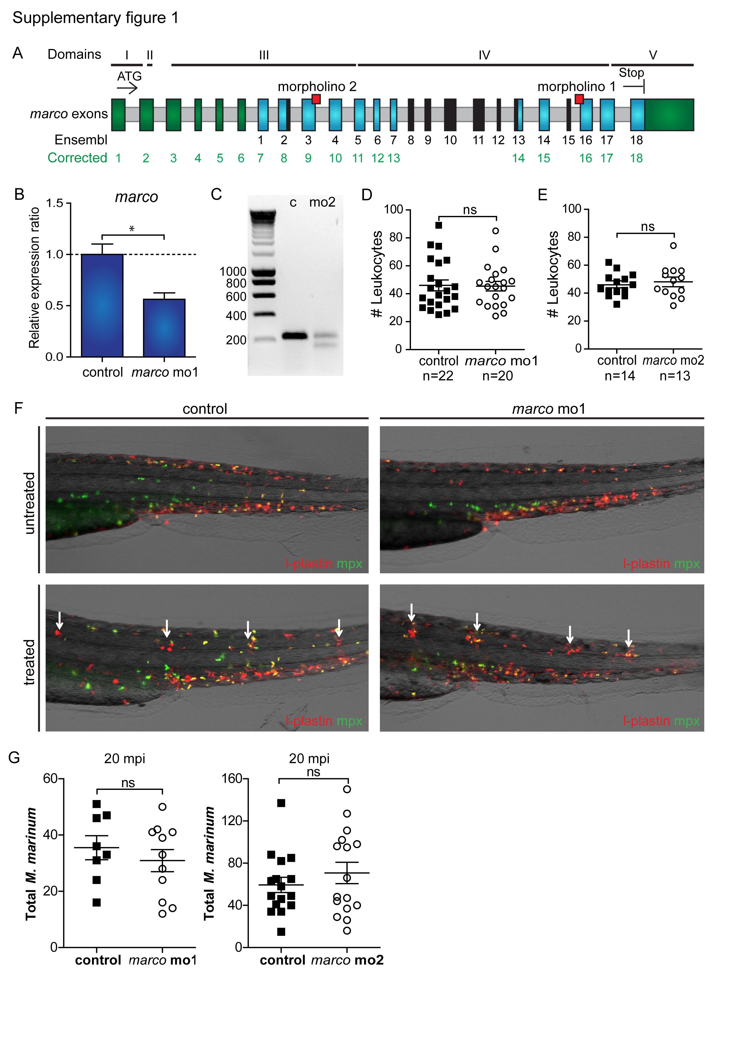
Validation of morpholino knockdown. (A) Corrected annotation of marco exons. The sequenced cDNA clone of marco shows that there are 6 additional exons (shown in green) prior to exon 1 in the current annotation in Ensembl (Zv9, ENSDARG00000059294), which was based on a predicted gene, LOC571584. Furthermore, the cDNA shows partial absence of Ensembl exons 2 and 13 and complete absence of exons 8–12 and 15 (shown in black), and an extended final exon (shown in green). Exons that are not different between the Ensembl annotation and the corrected annotation are shown in blue and introns are shown in grey. Exon–intron boundary sites targeted by morpholinos (red squares), the ATG and stop codon sites, and the conserved protein domains (I to V corresponding with Fig. 1) are indicated. (B,C) Knockdown of marco. The predicted effect of splice blocking morpholinos 1 is deletion of exon 16 in the collagenous domain and the predicted effect of morpholino 2 is deletion of exon 9 in the spacer domain. The exon deletion effect of morpholino 1 could not be confirmed by RT-PCR, but qPCR analysis (28 hpf) demonstrated that the marco mRNA level was effectively reduced (B). Deletion of exon 9 following knockdown of marco with morpholino 2 was confirmed by RT-PCR (C) using RNA from embryos injected with marco morpholino 2 (mo2) or control morpholino (c). The expected RTPCR product size for the control embryos is 228 bp and for marco mo2 morphants is 143 bp. (D,E) Normal leukocyte numbers in marco morphants. marco (D) morpholino 1 and (E) morpholino 2 morphants and control embryos were fixed at 30 hpf and were immuno-labelled with Ab against the general leukocyte marker L-plastin. Leukocyte numbers were counted blinded over the Duct of Cuvier, (ns = not significant). (F) Marco does not affect migration of leukocytes towards local inflammation sites. Representative images of copper sulphate treated control and marco morphant 3 dpf embryos. Embryos were immuno-labelled with Ab against the general leukocyte marker L-plastin (red signal) in combination with a neutrophil-specific Mpx TSA-staining (green signal) at 3 dpf. White arrows indicate accumulation of leukocytes at the local inflammation sites at the neuromasts. (G) Total number of M. marinum counted over Duct of Cuvier at 20 mpi for marco mo1 and mo2 morphants.