
|
Fig. S10
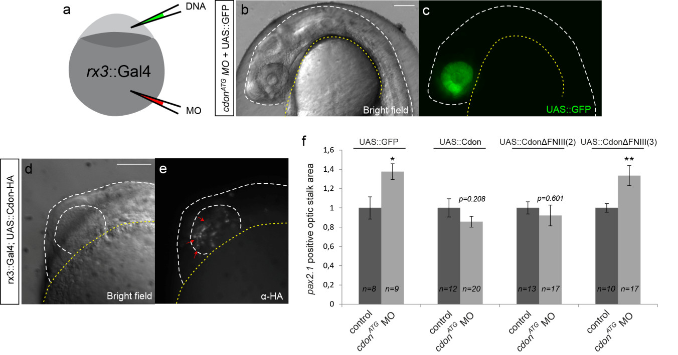
Overexpression of Cdon or CdonΔFnIII(2) in the eye rescues the optic stalk phenotype of cdon morphants. a) The schema depicts the injection method used in rescue experiments. Embryos expressing Gal4 protein in the rx3 positive domain (rx3::Gal4) were injected at 1-cell stage into the cell with UAS::GFP, UAS::Cdon, UAS::CdonΔFnIII(2) or UAS::CdonΔFnIII(3) DNA. After DNA injection, the eggs were injected with CdonATG MO into the yolk and the embryos were let develop until 26hpf (b). c) GFP expression was detected in the retinal and hypothalamic domains of the injected embryos. α-HA immunostaining in rx3::Gal4 embryos injected with UAS::Cdon DNA confirmed localized expression of Cdon in the retina of injected embryos (d, e). Note the membrane localization of the HA-signal (red arrows in e. f) Quantification of optic stalk pax2.1-positive domain in embryos co-injected with different UAS DNAs and cdonATG MO at 26-28hpf (*p<0.05, **p<0.01; Student’s t-test). The number of embryos analysed in each case is indicated in each column and are as follow: control/UAS::GFP, n=8; CdonATG MO/UAS::GFP, n=9; control/UAS::Cdon, n=12; CdonATG MO/UAS::Cdon, n=20; control/UAS::CdonΔFnIII(2), n=13; CdonATG MO/UAS::CdonΔFnIII(2), n=17; control/UAS::CdonΔFnIII(3), n=10; CdonATG MO/UAS::CdonΔFnIII(3), n=17. Error bars represent s.e.m. Note that the expansion of the pax2.1-positive optic stalk domain generated by cdonATG MO injection was less evident than that reported in Fig. 4l, likely owning to MO injection at two-four cells’ stage (see Methods). Scale bar: 100 μm.