
|
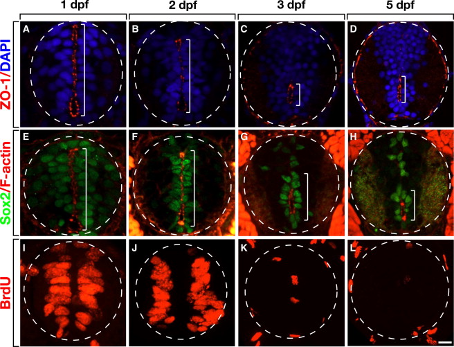
Fig. 1
Loss of Apical Polarity Correlates with Loss of Neural Precursors and Lumen Morphogenesis
All images show representative transverse sections at the level of the trunk spinal cord with dorsal up.
(A and B) At 1 and 2 dpf, ZO-1 is concentrated at apical membranes of cells lining a primitive lumen, which extends across the dorsoventral axis of the spinal cord (brackets).
(C and D) At 3 and 5 dpf, the primitive lumen is replaced with a ventrally positioned central canal marked by apically localized ZO-1.
(E–H) F-actin is similarly localized to apical membranes lining the primitive lumen and central canal. Additionally, most cells that express Sox2 are associated with F-actin localization.
(I–L) A BrdU pulse labels numerous cells lining the primitive lumen at 1 and 2 dpf, but at 3 and 5 dpf few cells incorporate BrdU. Scale bar equals 10 μm.
Reprinted from Developmental Cell, 27(4), Hudish, L.I., Blasky, A.J., and Appel, B., miR-219 Regulates Neural Precursor Differentiation by Direct Inhibition of Apical Par Polarity Proteins, 387-398, Copyright (2013) with permission from Elsevier. Full text @ Dev. Cell