
|
Fig. S4
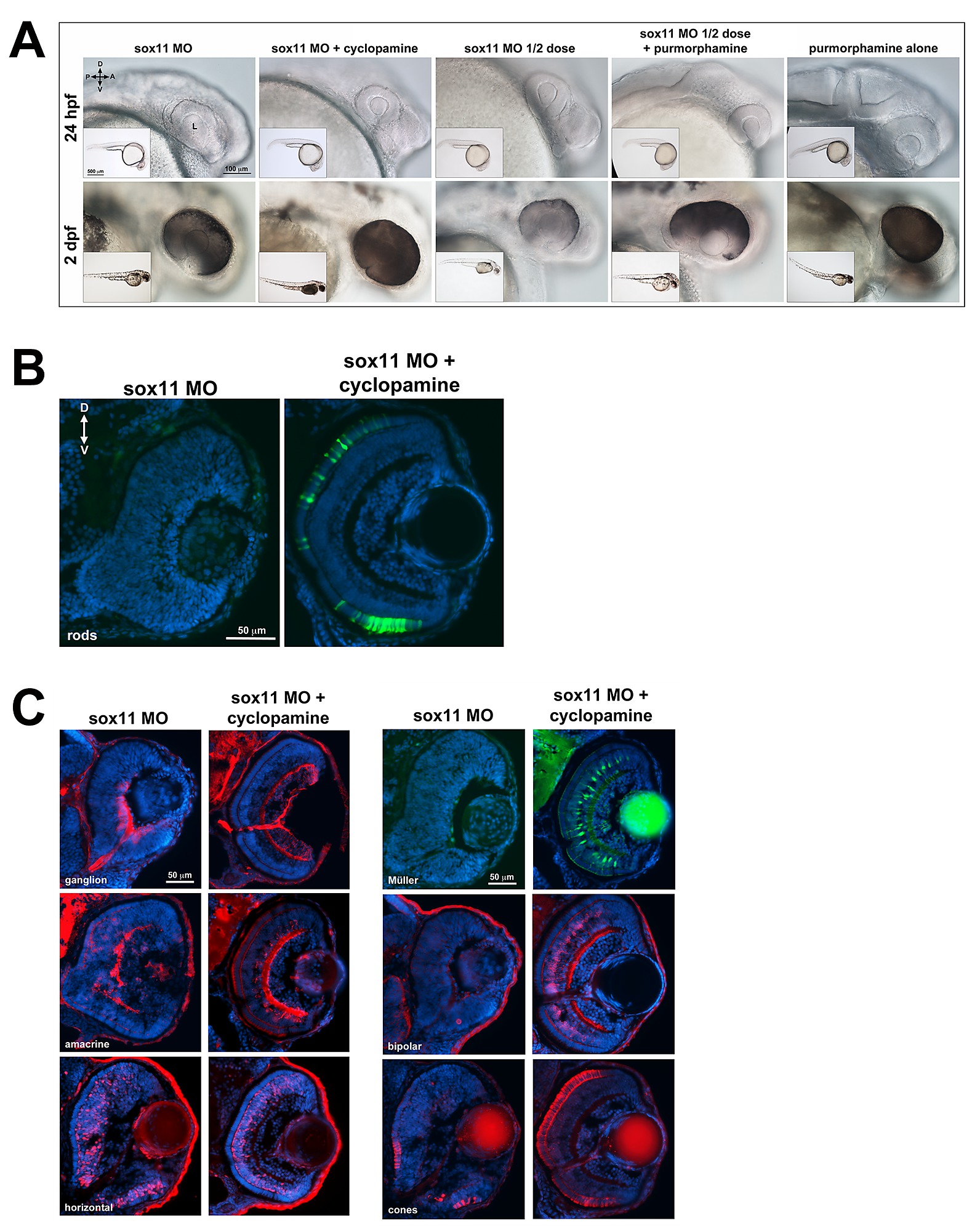
Elevated Hh signaling contributes to the abnormal ocular phenotypes displayed by sox11 morphants. (A) Representative brightfield images of sox11 morphants treated with cyclopamine, purmorphamine and their corresponding vehicle controls at 24 hpf and 2 dpf, taken from the set of embryos analyzed in Figure 5. Treatment with 75 uM purmorphamine alone did not cause any abnormalities (last column; 24 hpf control MO plus 75 μM purmorphamine alone, n = 123; 2 dpf control MO plus 75 μM purmorphamine alone, n = 114, 3 independent biological repeats). (B) Suppression of Hh pathway with cyclopamine rescued the rod photoreceptor defect in sox11 morphants at 3 dpf (right; number of embryos analyzed: sox11 MO, n = 20; sox11 MO + cyclopamine, n = 18; 3 independent repeats). (C) Retinal cell types were visualized by immunohistochemistry (ganglion, cones, amacrine, horizontal, and bipolar cells) or with a transgenic fluorescent reporter lines (Tg(gfap: GFP)mi2001) for Müller glia in sox11 morphants (left) and sox11 morphants treated with cyclopamine (right) at 3 dpf. The retinas of sox11 morphants treated with cyclopamine were well laminated and displayed normal distributions of all cell types (n = 15 per group, 3 independent repeats). D, dorsal; V, ventral; A, anterior; P, posterior; MO, morpholino; hpf, hours post fertilization; L, lens.