
|
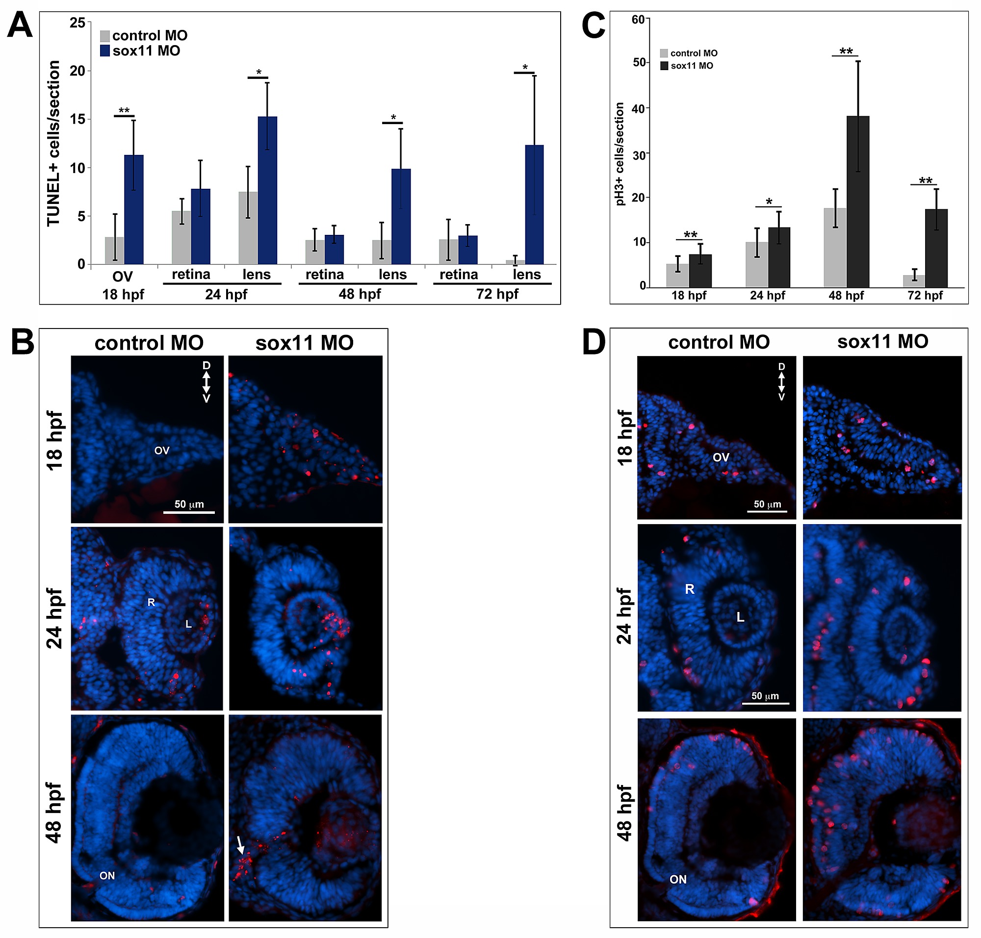
Fig. S2
Cell proliferation and apoptosis in sox11 morphants. (A) Quantification of TUNEL+ cells in the optic vesicle, lens, and retina of control and sox11 and morphants from 18–72 hpf. Sox11 morphants had an elevated number of TUNEL+ cells in the optic vesicle at 18 hpf. Additionally, sox11 morphants consistently displayed more TUNEL+ cells in the anterior lens compared to controls from 24 -72 hpf. Number of embryos analyzed: 18 hpf control MO, n = 20; 18 hpf sox11 MO, n = 22; 24 hpf control MO, n = 15; 24 hpf sox11 MO, n = 19; 48 hpf control MO, n = 10; 48 hpf sox11 MO, n = 13; 72 hpf control MO, n = 12; 72 hpf sox11 MO, n = 12; average of 3 independent biological replicates. **p<0.00001, *p<0.01, Student′s t-test. (B) Representative transverse sections of control (left column) and sox11 (right column) morphants at 18, 24, and 48 hpf, taken from the set of individuals analyzed in (A). At 48 hpf, TUNEL+ cells were detected within the colobomatous tissue and the region of the optic stalk in sox11 morphants (arrow, bottom right). (C) Sox11 morphant retinas had more PH3+ cells than controls from 18–72 hpf. Number of embryos analyzed: 18 hpf control MO, n = 12; 18 hpf sox11 MO, n = 15; 24 hpf control MO, n = 20; 24 hpf sox11 MO, n = 19; 48 hpf control MO, n = 10; 48 hpf sox11 MO, n = 12; 72 hpf control MO, n = 14; 72 hpf sox11 MO, n = 12; average of 3 independent biological replicates. **p<0.001, *p<0.01, Student′s t-test. (D) Representative transverse sections of control (left column) and sox11 (right column) morphants at 18, 24, and 48 hpf, taken from the set of individuals analyzed in (C). D, dorsal; V, ventral; MO, morpholino; hpf, hours post fertilization; ON; optic nerve; OV, optic vesicle; R, retina; L, lens.