
|
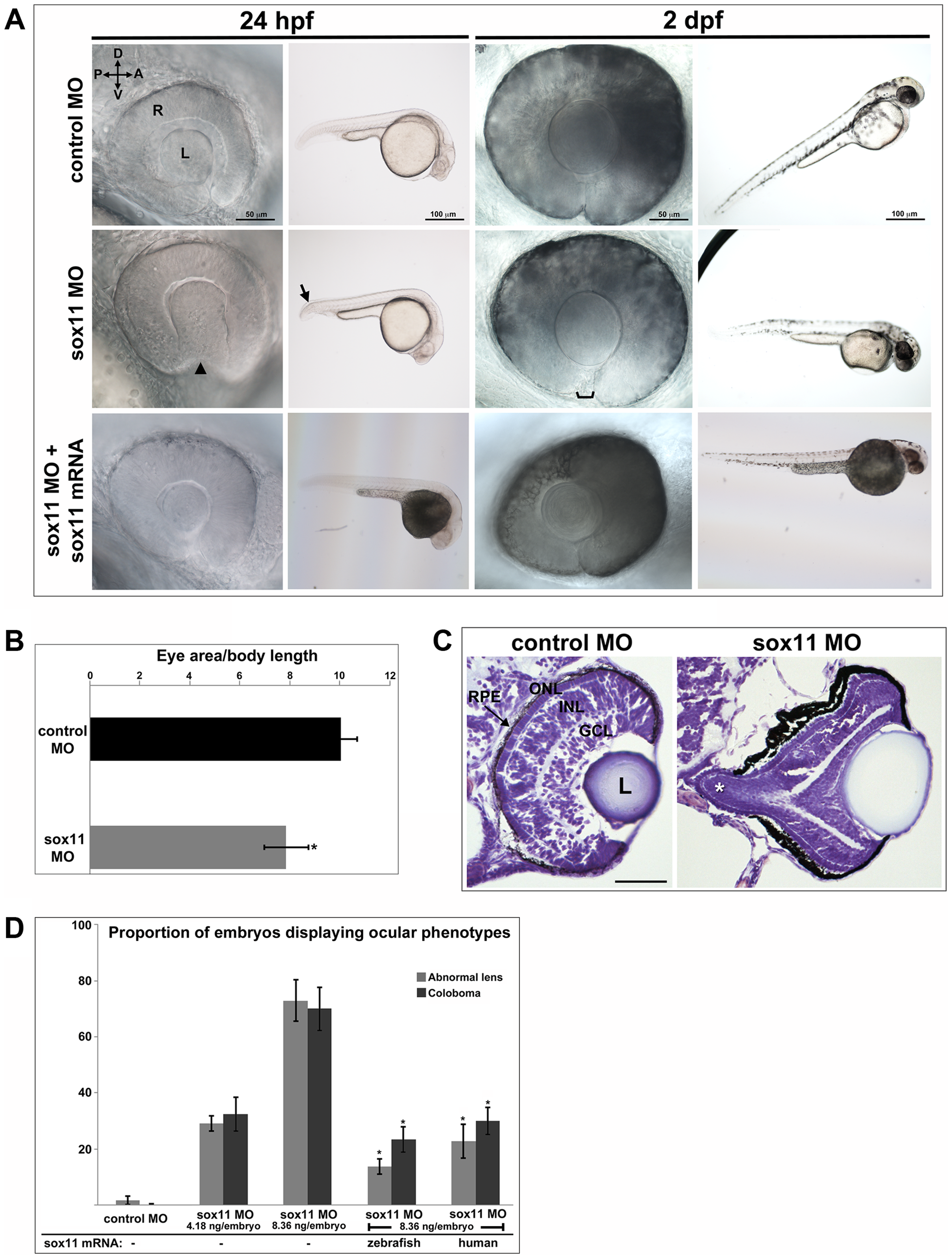
Fig. 2
Sox11 knockdown disrupts ocular morphogenesis and causes coloboma in zebrafish.
(A) Representative eye and body images of control and sox11 morphants (taken from the set of embryos analyzed in (D). At 24 hpf, approximately 70% of sox11 morphants displayed a malformed lens (arrowhead) and a posterior kink in the tail (arrow). At 2 dpf, a similar proportion of sox11 morphants displayed coloboma (bracket), and had a hypopigmented and underdeveloped ventral retina. Both the abnormal lens and coloboma phenotypes were rescued with co-injection of wild type zebrafish sox11 mRNA (bottom row). (B) Sox11 morphants were microphthalmic at 24 hpf. Eye area was normalized to body length (*p<0.0001, Student′s t-test; control MO: n = 10 embryos examined; sox11 MO: n = 12 embryos examined, 3 independent repeats). (C) Sections of 72 hpf control (left) and sox11 morphant eyes (right) stained with cresyl violet revealed the extrusion of the retina into the brain through the open choroid fissure of sox11 morphants (asterisk); n = 6 individuals examined per group. The thickened appearance of the dorsal RPE in the sox11 morphant retina is a staining artifact and was not observed in fresh tissue sections. Scale bar = 50 µm. (D) Injection of zebrafish and human sox11 mRNA rescued the ocular phenotypes in sox11 morphants. Number of embryos analyzed: 24 hpf control MO, 4.18 ng/embryo, n = 1007; 2 dpf control MO, 4.18 ng/embryo, n = 1001; 24 hpf sox11 MO, 4.18 ng/embryo, n = 309; 2 dpf sox11 MO, 4.18 ng/embryo, n = 294; 24 hpf sox11 MO, 8.36 ng/embryo, n = 559; 2 dpf sox11 MO, 8.36 ng/embryo, n = 392; 24 hpf sox11 MO 8.36 ng/embryo plus 2.0 ng/embryo zebrafish sox11 mRNA, n = 185; 2 dpf sox11 MO, 8.36 ng/embryo plus 2.0 ng/embryo zebrafish sox11 mRNA, n = 167; 24 hpf sox11 MO, 8.36 ng/embryo plus 0.3 ng/embryo human SOX11 mRNA, n = 130; 2 dpf sox11 MO, 8.36 ng/embryo plus 0.3 ng/embryo human SOX11 mRNA, n = 125. Three biological replicates were performed for all experiments. (*p<0.001, Student′s t- test). D, dorsal; V, ventral; A, anterior; P, posterior; L, lens; R, retina; hpf, hours post fertilization; dpf, days post fertilization; MO, morpholino; GCL, ganglion cell layer; INL, inner nuclear layer; ONL, outer nuclear layer; RPE, retinal pigmented epithelium.