
Image
|
Figure Caption
Fig. 3
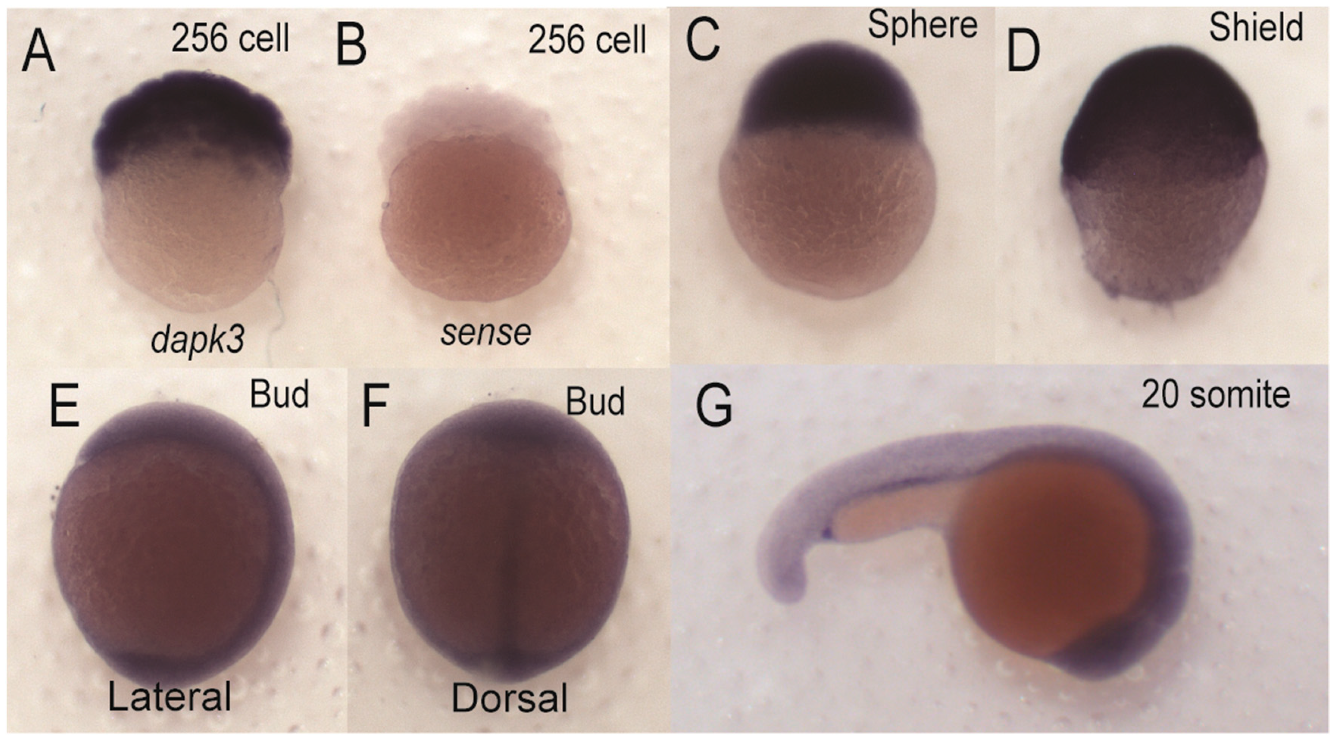
Spatial expression of zipk (dapk3) during early zebrafish development. Detection of zipk mRNA (dapk3) was carried out by whole-mount in situ hybridization using gene-specific probes on staged embryos at the 256 cells stage (A); sphere stage (C); shield stage (D); bud stage ((E) lateral view and (F) dorsal view); 20 somite stage (G); Negative control sense probes for zipk did not show staining at the 256 cell stage (B) or other stages (data not shown). Because of the difference in expression maternal stages (A–C) were developed for a shorter time than zygotic stages (D–G).
Figure Data
Acknowledgments
This image is the copyrighted work of the attributed author or publisher, and
ZFIN has permission only to display this image to its users.
Additional permissions should be obtained from the applicable author or publisher of the image.
Full text @ Int. J. Mol. Sci.