
|
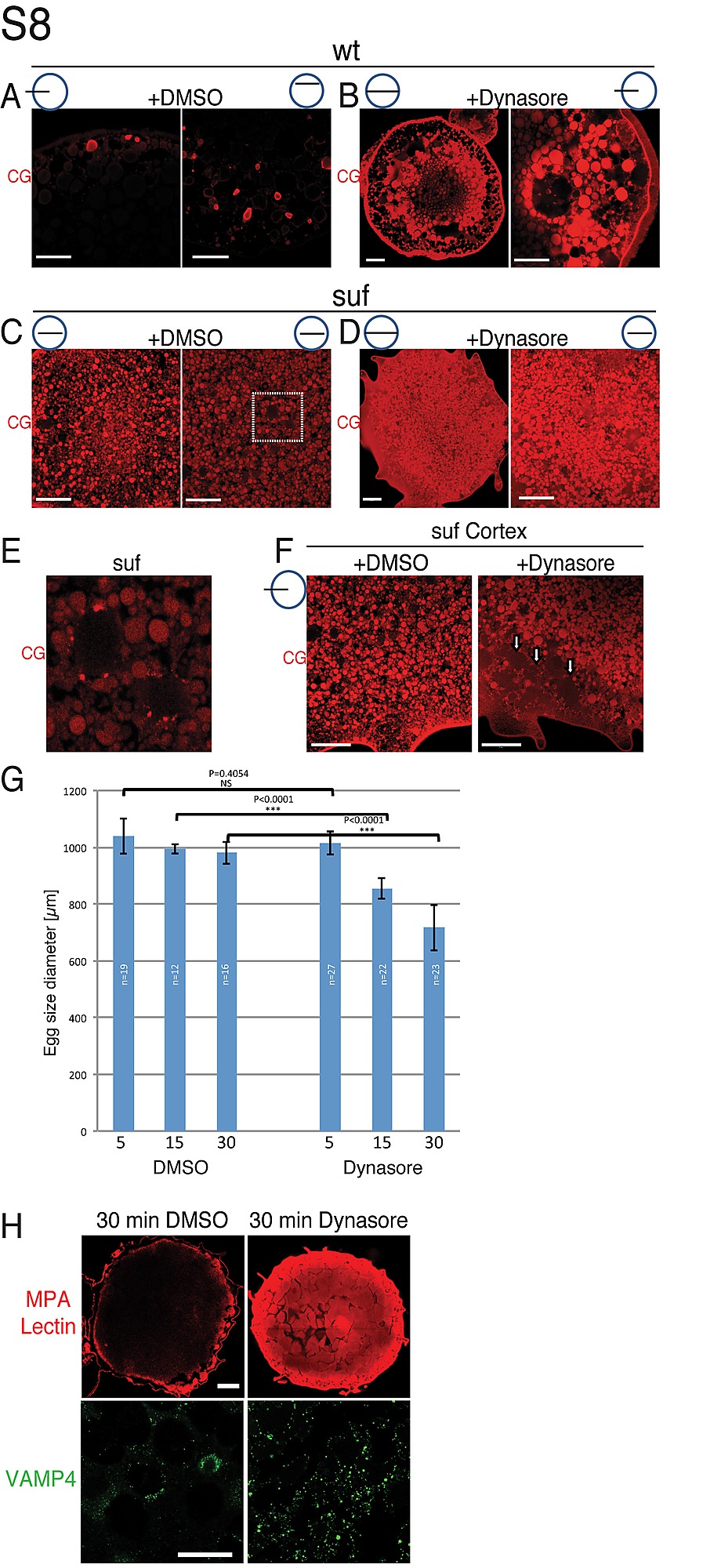
Fig. S8
The Dynamin inhibitor Dynasore mimics the suf/spastizin phenotype. Cortical granules stained with MPA-Lectin (red) in wild type (A, B) or suf/spastizin mutants (C, D) after treatment with the carrier DMSO (A, C) or with Dynasore (B, D). The dashed square in panel C indicates the magnification in panel E. (E) In untreated, suf/spastizin mutants MPA-Lectin (red) foci accumulated on the surface of MPA-negative vesicles similar to the cisternae discovered by EM suggesting that MPA-Lectin cargo is sorted, but not pinched off the compartment. (F) Cortex region of suf/spastizin oocyte before and after Dynasore treatment. Remarkably, suf mutants treated with Dynasore showed cortical granules with weak MPA-Lectin background (white arrowheads), which suggested that Dynasore inhibited sorting and removal of the MPA-Lectin cargo into buds. Small icon next to figures indicates the level of the optical section (black line) in the oocyte (blue circle). Scale bar: 50 μm. (G) Quantification of total egg size shown in Figure 7G. Note the significant size reduction of chorion elevation after 15 min. Error bars represent standard deviation. (H) Cellular marker analysis of ovulated stage V eggs. A 30 min treatment with Dynasore of ovulated eggs leads to an accumulation of MPA-lectin (red) and VAMP4 (green) similar to suf mutants. Scale bar: 50 μm.