
Image
|
Figure Caption
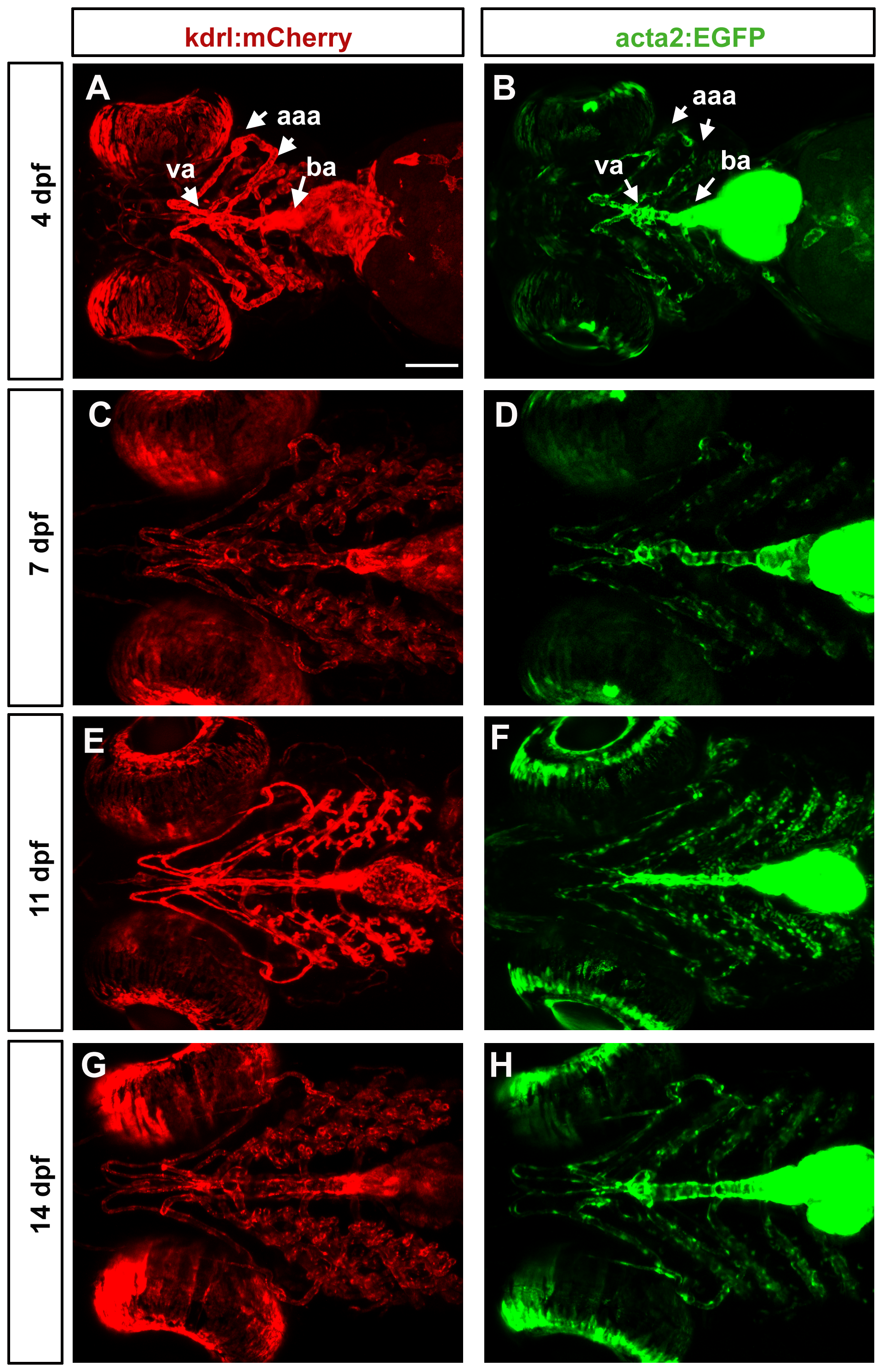
Fig. 4
Mural cell and endothelial development in the ventral head of larval zebrafish.
Confocal micrographs collected from a ventral point of view show a progressive increase in vessel complexity (red, A, C, E, G) and in density of mural cell coverage of aortic arch vessels (green, B, D, F, H) from 4 dpf (A, B), 7 dpf (C, D), 11 dpf (E, F) through 14 dpf (G, H). Heart expression of acta2:EGFP is maintained. aaa = aortic arch arteries; va = ventral aorta; ba = bulbus arteriosus. Scale bar in A represents 100 μm.
Acknowledgments
This image is the copyrighted work of the attributed author or publisher, and
ZFIN has permission only to display this image to its users.
Additional permissions should be obtained from the applicable author or publisher of the image.
Full text @ PLoS One