
|
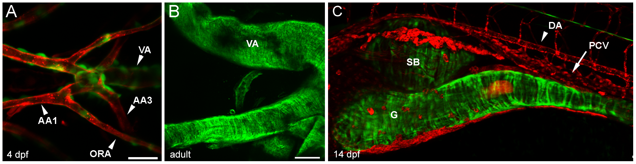
Fig. 2
Morphology of vascular and visceral mural cells in acta2:EGFP transgenic fish.
(A) Ventral pharyngeal region of a 4 dpf double transgenic Tg(acta2:EGFP)ca7; Tg(kdrl:mCherry)ci5 (mural cells are green and endothelial cells are red) zebrafish shows extensive mural cell coverage of the ventral aorta (VA) and lesser coverage on the smaller aortic arches (AA) or opercular artery (ORA). (B) Wholemount adult ventral aorta and attached afferent branchial arteries shows extensive smooth muscle coverage. (C) Lateral view of the gut (g) and swim bladder (b) of a 14 dpf double transgenic Tg(acta2:EGFP)ca7; Tg(kdrl:mCherry)ci5 zebrafish shows radial and circumferential smooth muscle on both gut and swim bladder, but sparse mural cells on the dorsal aorta (DA) and no visible cells on the posterior cardinal vein (PCV). Scale bar in A represents 25 μm. Scale bar in B and C represents 100 μm.