
|
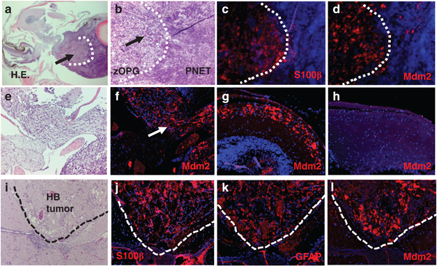
Fig. 7
Overexpression of Mdm2 in zOPGs and hindbrain tumor. (a) Hematoxylin and eosin (H&E) staining of a transverse section from an adult fish with a less affected eye and a gross eye tumor (arrow). (b) A fragmented line demarcates the zOPG with mild cellularity and a primitive neuroectodermal tumor (PNET) with high cellularity. Immunofluorescence revealed that only the zOPG exhibited S100β (c) and Mdm2 (d) overexpression, whereas the PNET did not. (e) H&E-stained transverse section showing the optic chiasm of the same fish. Mdm2 was only expressed in the optic nerve associated with the glioma (f, arrow). (g, h) High-magnification view of the two optic tectal lobes in (a) showing Mdm2 expression in the lobe associated with zOPG (g), but not in the contralateral lobe (h). (i) H&E staining of the hindbrain tumor. The tumor overexpressed radial glia markers S100β (j), GFAP (k) and Mdm2 (l).