
|
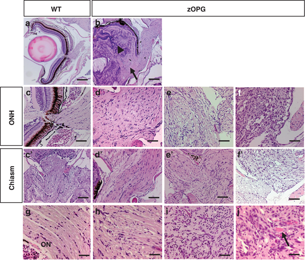
Fig. 4
Stable transgenic fish developed zOPGs. (a) Hematoxylin and eosin (H&E) staining of paraffin sections from a wild-type (WT) adult retina, and (b) an eye tumor showing coexistence of retinal dysplasia and ON hyperplasia. Note the high cellularity originating from the neural retina (arrowhead) and low cellularity derived from ON (arrow). (c) Enlarged view of normal ONH showing low cellularity, and transgenic fish show ON hyperplasia to neoplasia (d–f). (c2) Enlarged view of a normal optic chiasm showing low cellularity, and transgenic fish showing disorganization and increased cellularity in chiasms (d2–f2). (g) High-magnification view of a normal ON showing low cellularity, with zOPGs showing increased cellularity, nuclear atypia and vascular proliferation (h–j, arrow). Scale bars, 200μm for (a and b), 40μm for (c–f2) and 20μm for (g–j), respectively.