
|
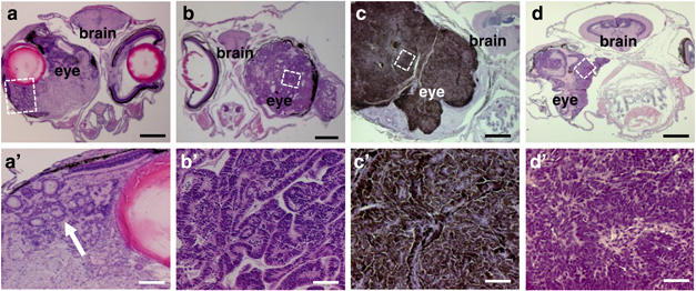
Fig. 3
Retinal tumors resulted from expression of Smoa1-EGFP in the stable transgenic line. (a, a2) Hematoxylin and eosin (H&E) staining of transverse section of a fish with retinal dysplasia; note the formation of rosette-like structures (arrow). (b, b2) An ocular tumor resembling medulloepithelioma with characteristic neural tube structures. (c, c2) A tumor showing features of pigmented ocular melanoma with heavy pigmentation and bland nuclei. (d, d2) An adult fish developed unilateral primitive neuroectodermal tumor (PNET). White frames indicate areas that were enlarged (not to scale). Scale bars, 400μm for (a–d), 40μm for (a2–d2), respectively.