
|
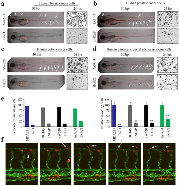
Fig. 1 Zebrafish faithfully report metastasis in human cancer cells. Confocal imaging (shown as z-stack images using 40 × magnification) shows that (a) metastatic MDA-MB-231 (MDA231) breast cancer cells (above) are dispersed throughout the fish body (arrows), whereas non-invasive T47D cells (below) remain in the confines of the yolk sac. These results are mirrored by the Transwell invasion assays, shown in each case to the right. Similarly (b) invasive DU145 prostate cancer cells show extensive spread throughout the fish compared with non-invasive LNCaP cells which do not. In colon cancer cells (c), highly invasive SW620 cells metastasize extensively compared with non-invasive HT29 cells. Relative invasion potential of pancreatic cancer cells (d) shows that more cells are distributed throughout the fish body for the relatively more invasive AsPC-1 cells compared with less invasive BxPC3 cells. Assessment of fish showing metastasis (see text) in each experiment compared with the invasion potential is shown in (e) showing the close correlation between relative invasion in vitro using the transwell assay and in vivo metastasis in fish. ** P > 0.001; * P >0.05. Using the Tg(kdrl:EGFP) transgenic zebrafish (f), cancer cells (red) that have spread throughout the vasculature (green) can be seen for MDA231 as show by successive imaging of 4 hours (200 × magnification). The presence of MDA231 cells that have extravasated into the body of the fish can clearly be seen (the white arrow tracks movement of a single cell and yellow arrow indicates movement of another).