
|
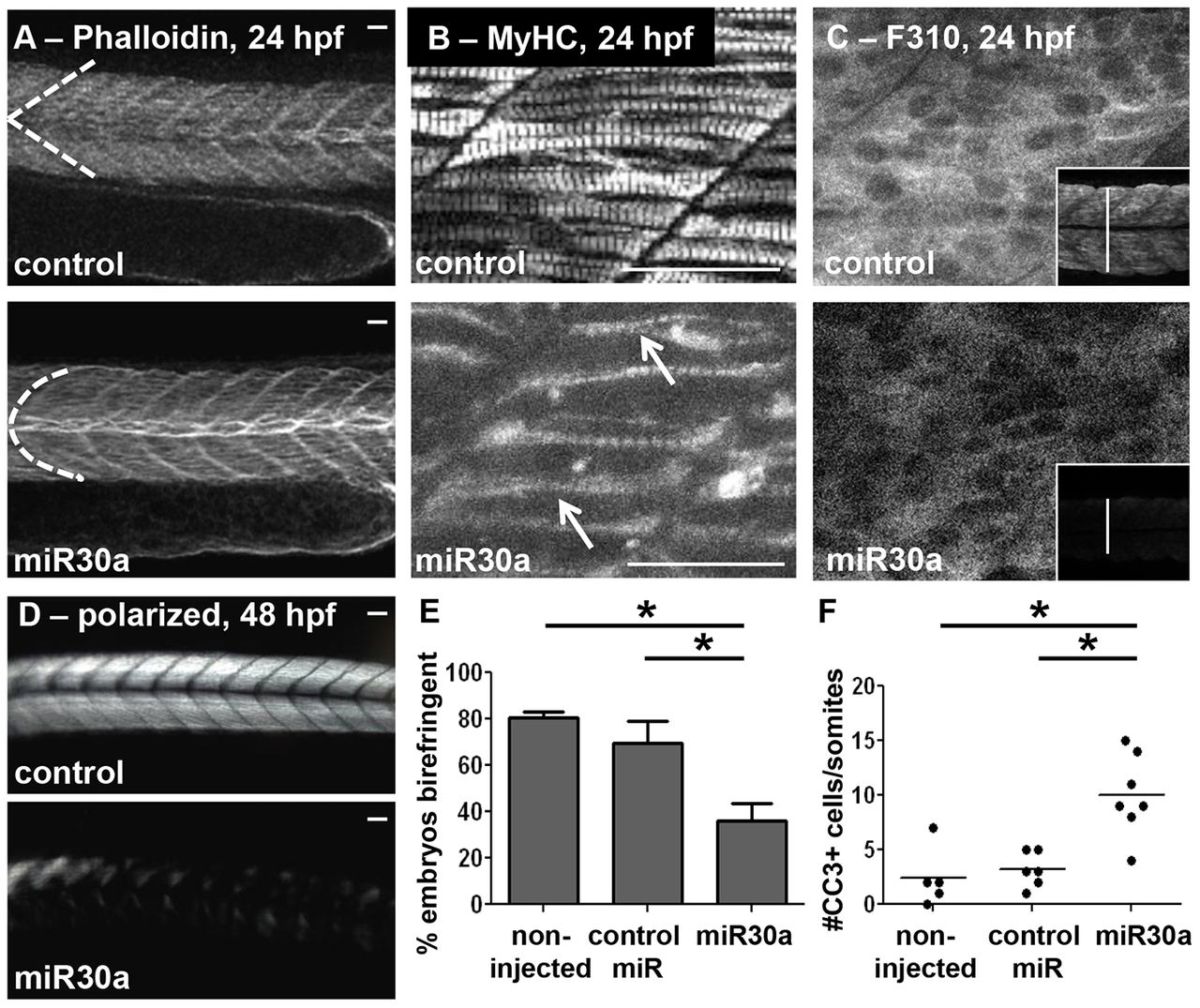
Fig. 3 miR30a overexpression functionally phenocopies six1a/b knockdown. Overexpression of miR30a leads to abnormal somite morphology as observed in (A) phalloidin-stained embryos (control, n = 42/49; miR30a, n = 46/77) at 24hpf, and confocal images of (B) both fast and slow MyHC with A4.1025 antibody (arrows indicate crossover of fibers) and (C) fast-muscle specific fibers with F310 antibody (the inset demonstrates a decrease in cross-sectional diameter of the trunk muscle) at 24 hpf. (D) Polarized-light-mediated detection of birefringence of embryos at 48 hpf revealed disarrayed muscle fiber organization upon miR30a overexpression. (E) A quantification (mean±s.e.m.) of the results shown in D. Non-injected, n = 23 total embryos; control miR, n = 25; miR30a, n = 21. *P = 0.008, ANOVA with Bonferroni′s post-hoc test. (F) A quantified increase in cleaved caspase-3 (CC3) is also observed upon miR30a overexpression at 24 hpf. Non-injected, n = 23 total embryos; control miR (1ng), n = 25; miR30a (0.5ng), n = 20. *P<0.05, ANOVA with Bonferroni′s post-hoc test. Scale bars: 50µm. For A, n = the number of embryos represented by each image/the total number of embryos analyzed.