
|
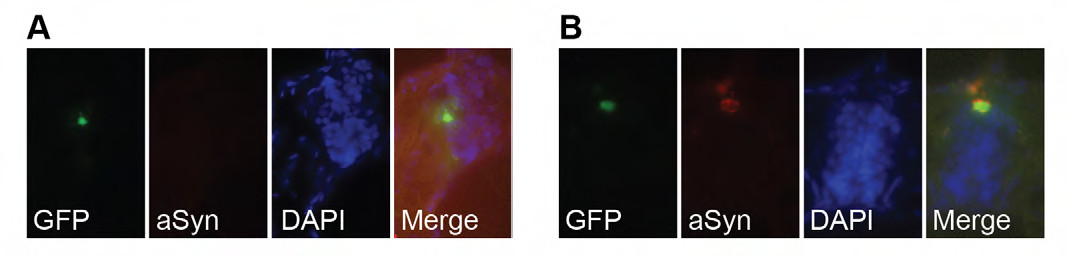
Fig. S1
Alpha-synuclein aggregates in zebrafish peripheral sensory neurons. Embryos were injected at the 1-cell stage with transgenes indicated in Fig. 1A. At 48 hours post-fertilization, embryos were fixed in 4% PFA, sectioned, and stained with anti-alpha-synuclein primary antibody, followed by Alexa 594-conjugated goat anti-mouse IgG secondary antibody. DAPI staining was used to visualize nuclei. (A,B) Cross-sections through the spinal cord of GFP- or aSyn-2A-GFP-expressing embryos. Green fluorescence indicates Rohon-Beard cells expressing the transgene. No aSyn staining was observed in control-injected cells (A). All aSyn-injected cells exhibited red aSyn staining (B).