
|
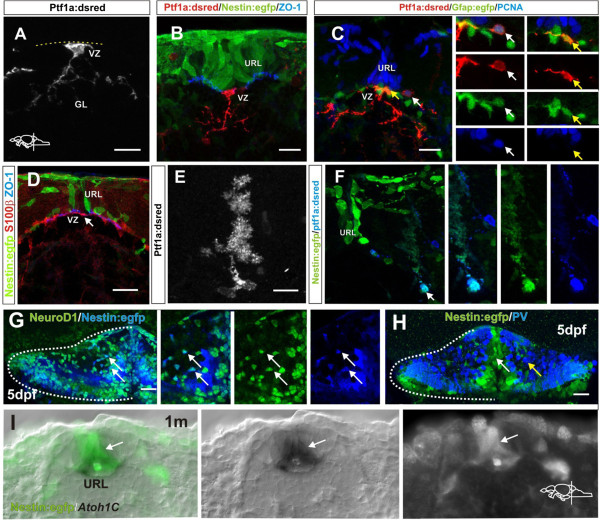
Fig. 6 Morphology of Ptf1a:DsRed cells in the VZ. (A) High magnification of a ventricularly located Ptf1a:DsRed+ cell with radial morphology in a juvenile zebrafish; (B) A Ptf1a:DsRed+ cell (red) with a radial morphology located at the ventricle, ventral to the Nestin:egfp+ cells (green) in the URL. Ventricle and apical junctions are outlined with ZO-1 (blue); (C) Ptf1a:DsRed+ and gfap:egfp+ cells (green) at the ventricle of the VZ. Proliferating cells in the progenitor niche are labeled with PCNA (blue) and glia are labeled by gfap:egfp (green). The white arrow depicts a proliferating Ptf1a:DsRed+and PCNA+ cell located lateral to the PCNA+ cells in the URL. The yellow arrow depict a horizontally oriented Ptf1a:DsRed+ and gfap:egfp+ cell at the ventricle; (D) Flat horizontally oriented s100β+ cells (red) are lining the VZ of the progenitor niche in adult zebrafish. The ventricle is outlined with the junctional marker ZO-1 (blue); (E) A Ptf1a:DsRed+ Bergmann glia in the adult cerebellum; (F) A Ptf1a:DsRed+ and Nestin:egfp+ Bergmann glia lateral to the progenitor niche (arrow); (G) Co-localization (arrows) of Nestin:egfp (blue) and NeuroD1 (green) in the cerebellum of a 5 day old larvae; (H) No overlap between Nestin:egfp (green, white arrow) cells and PV+ cells (blue, yellow arrow) is seen in the cerebellum of a 5-day-old larvae; (I) Overlapping expression of Atoh1c and Nestin:egfp in the URL. C: Cerebellum, GL: Granule cell layer, URL: Upper rhombic lip, VZ: Ventricular zone.