
|
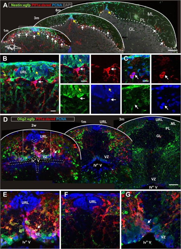
Fig. 3 Proliferative activity of cerebellar progenitors diminishes during juvenile stages. (A) Confocal maximum projection of cerebellar cross sections at a similar level of juvenile, young and adult cerebellum. Proliferating cells are labeled with proliferating cell nuclear antigen (PCNA) in blue. Nestin:egfp+ cells are green, Ptf1a:DsRed+ cells are red and the general tissue morphology is depicted with DAPI staining in grey. Yellow arrows depict the distinct niche in the URL where proliferating Nestin:egfp+/PCNA+ progenitors are located. White arrows depict Ptf1a:DsRed+ cells that localize to the ventral part of the IVth ventricle and to the cerebellar parenchyma. The majority of the parenchymal Ptf1a:DsRed+ cells are PCNA- while most of the Ptf1a:DsRed+ cells at the VZ close to the ventricle are PCNA+. (B-C) High magnification single confocal planes of the boxed areas in A. (B) A proliferating Ptf1a:DsRed+ /PCNA+ progenitor at the IVth ventricle (white arrow) and many proliferating Nestin:egfp+/PCNA+ cells in the URL (yellow arrow); (C) A proliferating Ptf1a:DsRed+ cell in the cerebellar parenchyma (white arrow); (D) Confocal maximum projections of juvenile and adult cerebellar cross sections showing a decline in VZ progenitor activity. (E) High magnification of boxed area in D. Proliferating Ptf1a:DsRed+ (red) and Olig2:egfp+ (green) cells are found in VZ and the cerebellar parenchyma in a two-week-old juvenile fish. Note the absence of Ptf1a:DsRed+ and Olig2:egfp+ cells among the PCNA+ cells in the URL. The white arrow depicts the Ptf1a:DsRed+ and Olig2:egfp+cells laminating from the ventricular surface into the parenchyma; (F) Ptf1a:DsRed+ cells adjacent to proliferating PCNA+ cells in a one-month-old fish; (G) Proliferating Ptf1a:DsRed+ and Olig2:egfp+ in the VZ of the fourth ventricle (arrow) in a one-month-old fish. The white arrow shows the Ptf1a:DsRed+ and Olig2:egfp+ cells laminating from the ventricular surface into the parenchyma.