
|
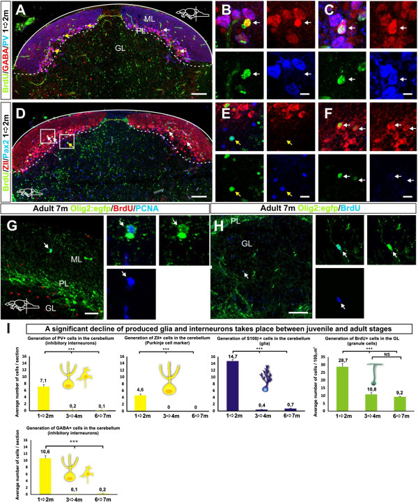
Fig. 8 Generation of different cerebellar cell types. (A) Confocal maximum projection of a cerebellar cross section in a juvenile zebrafish showing BrdU labeled GABA+ inhibitory neurons (white arrows) and BrdU+/GABA+/PV+ neurons (yellow arrows); (B) High magnification of a single confocal plane showing a neuron co-localizing with BrdU and GABA (white arrow); (C) High magnification of a single confocal plane showing a neuron co-localizing with BrdU, GABA and PV (white arrow); (D) Confocal maximum projection of a cerebellar cross section showing BrdU labeled ZII+ Purkinje neurons (white arrows) and a Pax2+ Golgi neuron (yellow arrow); (E) High magnification of a single confocal plane showing a Golgi neuron co-localizing with BrdU and Pax2 (white arrow); (F) Magnified single confocal plane showing Purkinje neurons co-localizing with BrdU and ZII (white arrows); (G) A rare proliferating oligodendrocyte progenitor (PCNA/Olig2:egfp+) detected in the cerebellar parenchyma (arrow); (H) A putative oligodendrocyte labeled with BrdU+and Olig2:egfp+ detected in the brain parenchyma six weeks after the BrdU pulse; (I) Quantifications of the cell types produced in the cerebellum of juvenile and adult zebrafish after four weeks BrdU pulse chasing. A significant decline of cerebellar inhibitory neuron and glia production between juvenile and adult stages is detected while granule cell production declines during juvenile stages but is still maintained at a high level in the adult zebrafish (n = 5, P <0.001).