
|
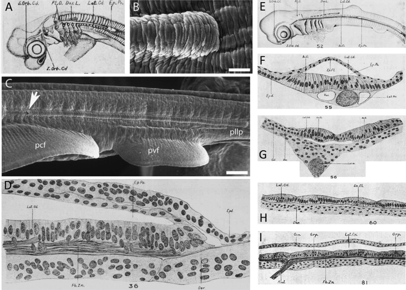
Fig. 6 In cartilaginous fishes, an epidermal pocket or tunnel forms above the posterior lateral line primordium as it elongates. (A) Lateral surface view of a 20 mm spiny dogfish (Squalus acanthias) specimen. The elongating posterior lateral line primordium (lateral sensory cord/ridge, Lat.Cd.) and epidermal pocket or tunnel (Ep.Po.) are clearly visible (sketch reproduced from Johnson, 1917). (B) Scanning electron micrograph of the advancing tip of the posterior lateral line primordium within the epidermal pocket/tunnel in the little skate Leucoraja erinacea (reproduced from Gillis et al., 2012). (C) The sensory ridge extends caudally along the entire length of the trunk. The opening to the epidermal tunnel is indicated by a white arrow (reproduced from Gillis et al., 2012). (D) Horizontal section through the growing end of the posterior lateral line primordium in a 15 mm S. acanthias specimen, showing the double-layered epidermal pocket/tunnel (Ep.Po) forming dorsal to it (modified from Johnson, 1917). (E) Lateral surface view of a 27 mm S. acanthias specimen (reproduced from Johnson, 1917). (F) Transverse section through a 27 mm S. acanthias specimen at a level caudal to the first dorsal fin, where the lateral sensory ridge is still covered by the superficial epithelial pocket (reproduced from Johnson, 1917). (G) Transverse section through a 27 mm S. acanthias specimen at a level rostral to the epidermal pocket/tunnel, where the lateral sensory ridge is exposed to the surface (reproduced from Johnson, 1917). (H) Horizontal section through a 29 mm S. acanthias specimen. The hair cells are arranged into small groups (reproduced from Johnson, 1917). (I) Longitudinal section of the lateral canal in a S. acanthias pup. The sketch shows the general histological morphology of the sensory epithelium, which is now enclosed within an epithelial canal (Lat.Cn.), and its innervation (reproduced from Johnson, 1917). Abbreviations: Ac.O., accessory organ; Can., canal wall; Der, dermis; Dor.L., anlage of dorsal line of pit organs; Ep.Fl., epidermal fold; Ep.Po., epidermal pocket or tunnel; Epd., epidermis; Fb.Zn., longitudinal nerve fiber zone; Grp., group of hair cells; I.Orb.Cd., supraorbital sensory cord; Lat.Cd., lateral sensory cord; Lat.Cn., lateral canal; Lat.Nv., lateral line nerve; pcf, pectoral fin; pllp, posterior lateral line primordium; Pt.O., anlage of isolated pit organs; pvf, pelvic fin; Rml., ramulus lateralis (little branch of lateral line nerve); S.Orb.Cd., supraorbital sensory cord; Sn.Cl., secondary sense cell; Vas., vascular space.
Reprinted from Developmental Biology, 389, Piotrowski, T., Baker, C.V., The development of lateral line placodes: taking a broader view, 68-81, Copyright (2014) with permission from Elsevier. Full text @ Dev. Biol.