
|
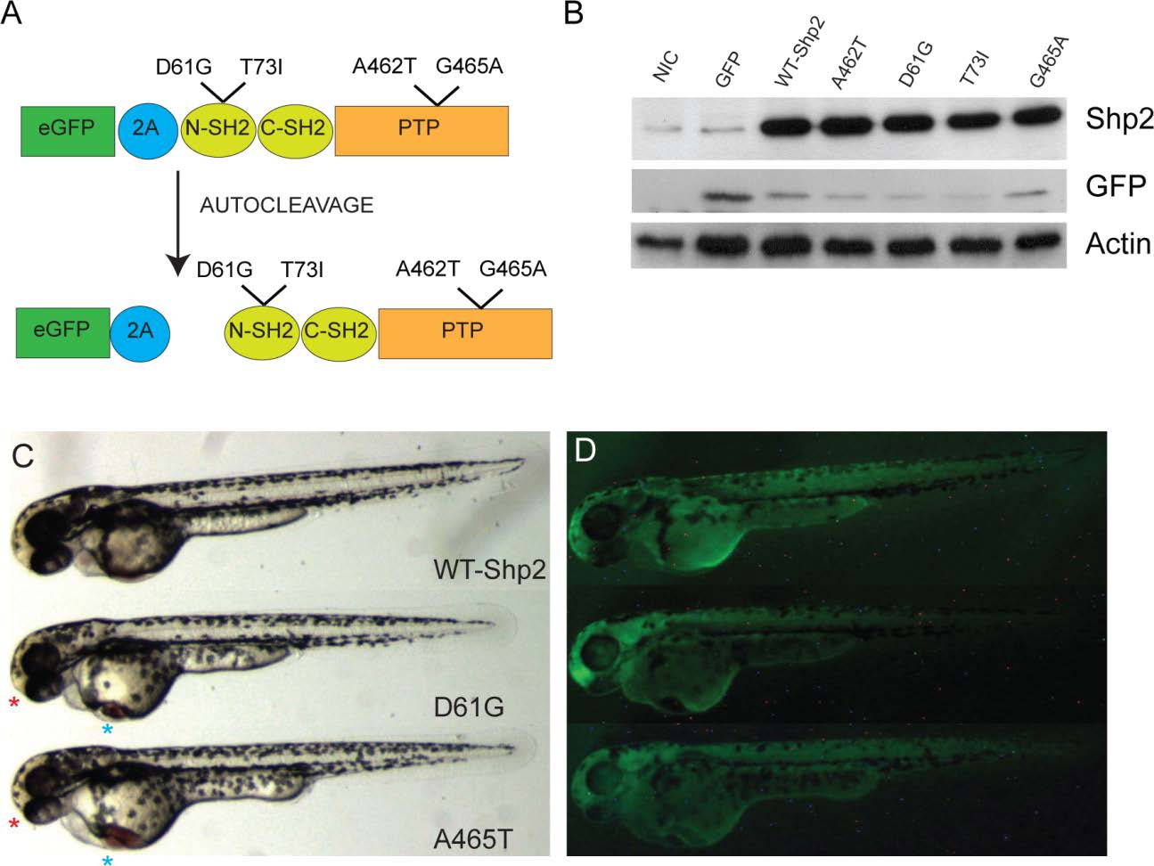
Fig. S1 EGFP-Peptide 2A-Shp2 fusions induced defects in zebrafish embryos. (A) Schematic representation of eGFP-Peptide 2A-Shp2 with D61G, T73I (NS) and A462T, G465A (LS) mutations indicated. The full length fusion protein is produced and is then cleaved autoproteolytically, resulting in eGFP and (mutant) Shp2. (B) One- or two-cell stage embryos were injected with in vitro transcribed mRNAs encoding the indicated proteins. Immunoblotting analysis reveals that autoproteolytic cleavage of the fusion proteins is highly efficient in zebrafish embryos. The injected embryos were lyzed at 10 hpf, proteins were separated on SDS-polyacrylamide gels, blotted and probed with antibodies specific for Shp2, GFP and β-actin as a loading control. (C-D) Two representative injected embryos (D61G and A462T) show craniofacial defects (red asterisks), a decrease in body length and cardiac edema (blue asterisks). These defects are absent in WT-Shp2 injected control embryos. Injection efficiency was checked by fluorescence microscopy with a GFP filter.