
|
Fig. 4
Asymmetric Responses of left dHb Neurons to Light Are Dependent upon the Eyes
(A and B) Dorsal views of live wild-type (A) and chkne2611 mutant (B) fish at 4 dpf.
(C) Dorsal view of the dHb nuclei in a chkne2611 mutant showing that mitral cells inputs remain asymmetrically directed to the right dHb.
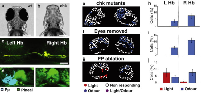
(D) Dorsal views of the epithalamus of a Tg(elavl3:GCaMP5G) 3 dpf fish prior (left) and 4 dpf fish subsequent (right) to ablation of the parapineal (pseudocolored blue).
(E–G) Images of color-coded dHb neuron responses to light (red) and odor (blue) in single z plane of a 4 dpf Tg(foxD3:GFP × lhx2a:Gap43-YFP) chkne2611 mutant fish (E), a fish in which eyes were surgically removed at 1 dpf (F), and a Tg(foxD3:GFP × elavl3:GCaMP5G) embryo with late ablation of the parapineal (G).
(H–J) Bar graphs showing average dHb neuron responses to light and odor for chk mutant (H) eye-ablated (I) and parapineal-ablated (J) fish. Fish without eyes show loss of dHb visual responses, whereas fish without a parapineal still show visual responses. For chkne2611 mutant and eye-ablated fish, an average of 12% (n = 6 fish) and 16% (n = 6 fish), respectively, of neurons responded to odor. For late parapineal-ablated fish (n = 3 fish), dHb neurons show an asymmetric distribution of responses to light (9% of neurons) and odor (11% of neurons) comparable with that of the wild-type. Error bars indicate the SEM.
Scale bars, 20 μm. Hb, habenulae; Pp, parapineal; LHb, left habenula nucleus; R Hb, right habenula nucleus.