
|
Fig. 1
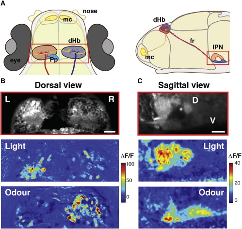
Responses to Visual and Odor Stimuli Are Lateralized in the dHb and Segregated Dorsoventrally in the IPN
(A) Schematic dorsal and sagittal views of 4 days postfertilization (dpf) zebrafish showing left (red) and right (blue) dHb nuclei and their asymmetric afferents from olfactory mitral cells (yellow) arborizing in the right dHb nucleus and parapineal neurons (cyan) arborizing in the left dHb nucleus. Neurons of the left dHb predominantly innervate the dorsal IPN, while neurons of the right dHb innervate the ventral IPN.
(B) Example of a two-photon image of a single z plane of the dHb (14 μm below the skin) of a Tg(elavl3:GCaMP5G) 4 dpf fish (top) and corresponding color-coded calcium signals that are LR lateralized in dHb neurons in response to a nonlateralized presentation of light (middle) and odor (bottom) stimuli. Each panel is an average of two stimulus trials. The relative change in fluorescence (ΔF/F) is expressed as a percentage.
(C) Example of a two-photon image of a lateral view of the IPN of a Tg(elavl3:GCaMP5G) 4 dpf fish (top) and corresponding color-coded calcium responses to light (middle) and odor (bottom). Each panel is an average of three trails. Responses to light are predominantly localized in the dorsal IPN, while those to odor are localized in the ventral IPN. Note that the dorsal IPN has a higher basal fluorescence (as does the left dHb nucleus).
Scale bars, 20 μm. D, dorsal; V, ventral; fr, fasciculus retroflexus; IPN, interpeduncular nucleus; dHb, dorsal habenulae; L, left; R, right; mc, mitral cells; Pp, parapineal. See also Figure S1.