
|
Fig. 4
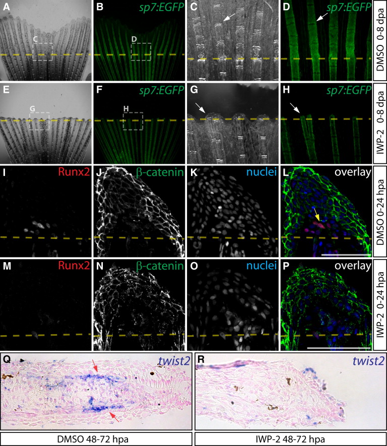
Wnt/β-Catenin Is Required for Osteoblast EMT and Dedifferentiation
(A–H) Regeneration of Tg(sp7:EGFP) fins after treatment with DMSO (A–D) or IWP-2 (E–H, 10 μM from 0 to 8 dpa). Rotterman contrast (A, C, E, G) and epifluorescence (B, D, F, H) images show sp7:EGFP expression in osteoblasts (white arrows) before amputation (A, B, E, F) and at 8 dpa (C, D, G, H). Shown are 25× images from one of three fish for control and IWP-2 groups and regions within dashed white boxes are shown at 120× magnification.
(I–P) Immunostaining for Runx2 (red) and β-catenin (green) on 24 hpa sections from fish exposed to DMSO (I–L) or Wnt inhibitor (M–P, 10 μM IWP-2 from 0 to 24 hpa). The yellow arrow indicates Runx2+ cells with nuclear-localized β-catenin. Nuclei are stained blue. Scale bars represent 50 μm.
(Q and R) twist2 in situ hybridization on fins from DMSO (Q) and IWP-2-treated (R, 10 μM from 48 to 72 hpa) fish harvested 72 hpa. The red arrows point to twist2-expressing osteoblasts.